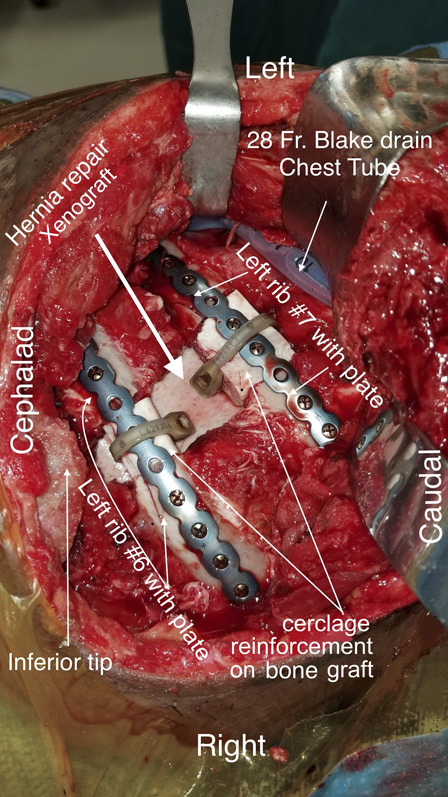
Fig. 13.

Intraoperative photograph showing repair of a traumatic hernia with destruction of a portion of the intercostal muscles between ribs 5/6, 6/7, and 7/8. Bone deficit is seen in the left sixth and seventh ribs. A thick white arrow shows a xenograft underlay repair of the traumatic hernia. Ribs 6 and 7 are repaired with precontoured rib-specific plates and a cadaveric bone graft to bridge the gaps. Polymer cerclage bands are utilized to further secure the bone graft to the plates when there is an insufficient number of anchoring screws (≤3). The inferior tip of the scapula is shown and labeled. A 28 French gauge Blake drain is used as a chest tube.
