Figure 1.

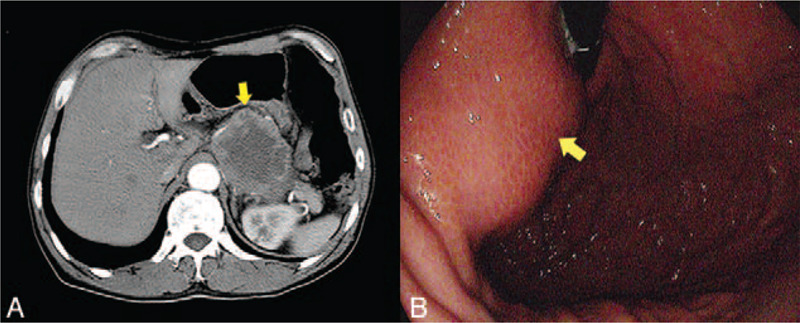
Images of CT and gastroscopy. Contrast-enhanced CT showed a large exophytic mass (yellow arrow) on the body of the stomach (A). The gastroscopy indicated a submucosal mass (yellow arrow) (B).

Images of CT and gastroscopy. Contrast-enhanced CT showed a large exophytic mass (yellow arrow) on the body of the stomach (A). The gastroscopy indicated a submucosal mass (yellow arrow) (B).