Figure 2.

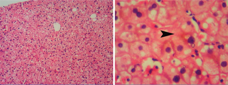
Microscopic features of the liver biopsy specimens. A: Normal hepatic lobule (hematoxylin and eosin staining, original magnification ×100). B: Bland cholestasis (hematoxylin and eosin staining, original magnification ×400).

Microscopic features of the liver biopsy specimens. A: Normal hepatic lobule (hematoxylin and eosin staining, original magnification ×100). B: Bland cholestasis (hematoxylin and eosin staining, original magnification ×400).