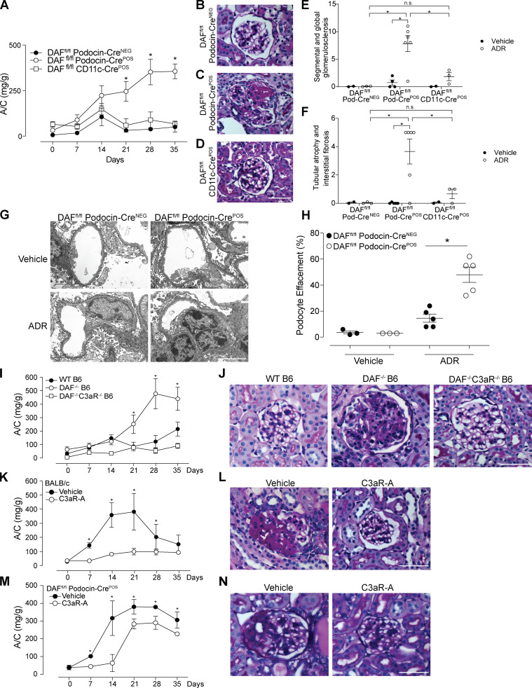
Figure 2.
Podocyte-specific removal of DAF from podocytes increases susceptibility to ADR-induced injury through C3a/C3aR signaling. (A–F) Urinary A/C at weekly intervals (A) and representative renal histological (PAS) lesions (B–D) with data quantification (E and F) of male DAFfl/fl podocin-CreNEG (n = 10), DAFfl/fl podocin-CrePOS (n = 19), and DAFfl/fl CD11c-CrePOS (n = 5) mice injected with ADR (20 mg/kg, i.v.) and sacrificed after 5 wk. (G and H) Representative electron micrographs (G) and quantification (H) of podocyte effacement in 8-wk-old DAFfl/fl podocin-CrePOS or DAFfl/fl podocin-CreNEG mice at 5 wk after treatment with saline or ADR (20 mg/kg, i.v.). (I and J) Urinary A/C at weekly intervals (I) and representative renal histological (PAS) lesions (J) of WT (n = 16), DAF−/− (n = 13), and DAF−/−C3aR−/− (n = 4) male B6 mice injected with ADR (20 mg/kg, i.v.). (K and L) Urinary A/C at weekly intervals (K) and representative renal histological (PAS) lesions (L) of male BALB/c mice given ADR (10 mg/kg, i.v.) and treated with C3aR-A (1 mg/kg/d, s.c.; n = 5) or saline (n = 5). (M and N) Urinary A/C at weekly intervals (M) and representative renal histological (PAS) lesions (N) of male B6 DAFfl/fl podocin-CrePOS mice given ADR (20 mg/kg, i.v.) and treated with C3aR-A (1 mg/kg/d, s.c.; n = 5) or saline (n = 5). *P < 0.05 versus podocin-CreNEG, WT, or C3aR-A. All experimental data were verified in at least three independent experiments. n.s., not significant. Scale bars: 50 µm. Error bars are SEM.

