Figure 7.
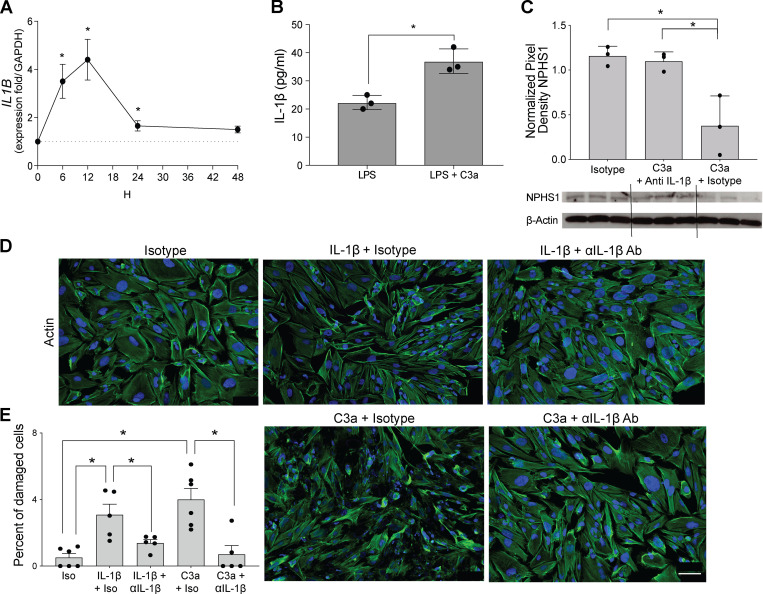
IL-1β mediates complement-induced podocyte injury in vitro. (A) IL1B gene expression in hiPod at serial time points after C3a stimulation (50 nM). (B) IL-1β levels in the supernatants of hiPod at 24 h after LPS (5 ng/ml) with or without C3a stimulation. (C) Nephrin expression in hiPod at 24 h after stimulation with isotype, C3a + anti–IL-1β–neutralizing antibody, and C3a + isotype (WB). *P < 0.05 versus 0 h. (D and E) Representative images (D) and quantification (E) of cell injury of hiPod exposed to isotype control, IL-1β (50 ng/ml) + isotype control, or IL-1β (50 ng/ml) + anti–IL-1β–neutralizing antibody (0.5 µg/ml; upper row). In the bottom row, the same cells were exposed to C3a + isotype control or C3a + anti–IL-1β–neutralizing antibody for 1 h. All experimental data were verified in at least two independent experiments. *P < 0.05. Scale bars: 100 µm. Error bars are SEM.

