Figure 8.
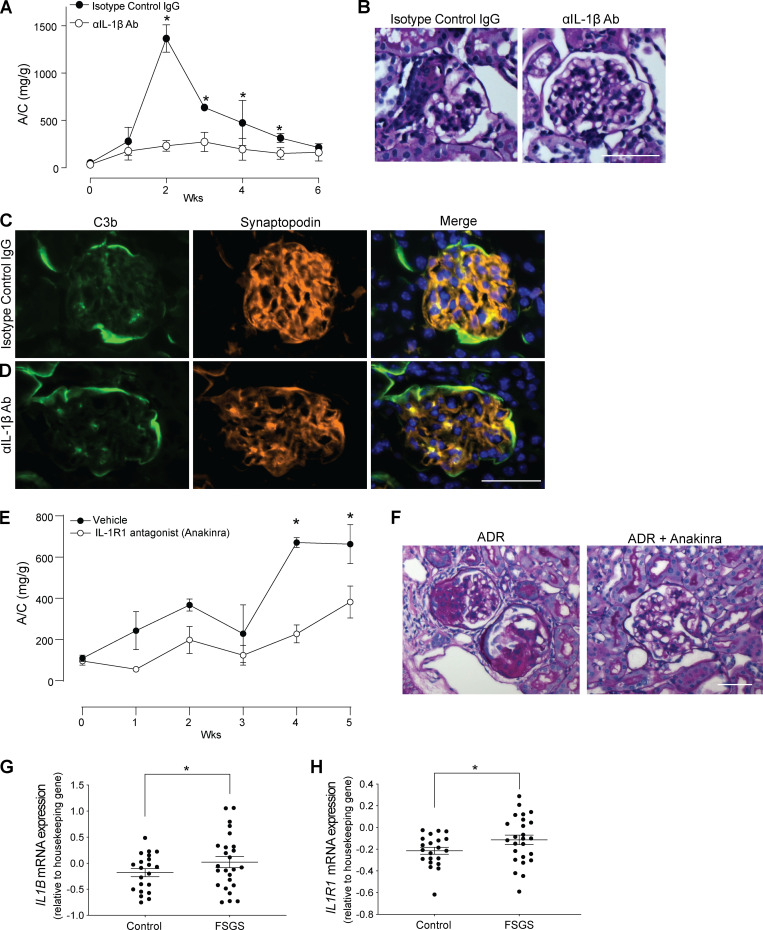
IL-1β mediates complement-induced podocyte injury in vivo. (A and B) Urinary A/C (A) and representative renal histological changes (B) in BALB/c mice injected with 10 mg/kg of ADR (i.v.) and rat anti-mouse IL-1β–neutralizing mAb (50 µg/mice twice per week, i.p.; n = 4) or isotype control (n = 4). Scale bars: 50 µm. *P < 0.05 versus anti–IL-1β–neutralizing antibody at the same time point. (C and D) Representative images of C3b deposition in the glomeruli of mice treated with (C) anti–IL-1β–neutralizing antibody or (D) isotype control. (E and F) Urinary A/C (E) and representative renal histological changes (F) in B6 DAFfl/fl podocin-CrePOS male mice injected with 10 mg/kg of ADR (i.v.) and anakinra (25 mg/kg/d through s.c. pumps; n = 4) or vehicle control (n = 3). Scale bars: 50 µm. *P < 0.05 versus anakinra at the same time point. (G and H) IL1B (G) and IL1R1 mRNA expression (H) in glomeruli of human biopsy specimens with pathological diagnosis of FSGS compared with normal kidneys. Data are from previously published microarray studies by Ju et al. (2013) and were subjected to further analysis using Nephroseq. *P < 0.05. All experimental data were verified in at least two independent experiments. Error bars are SEM.

