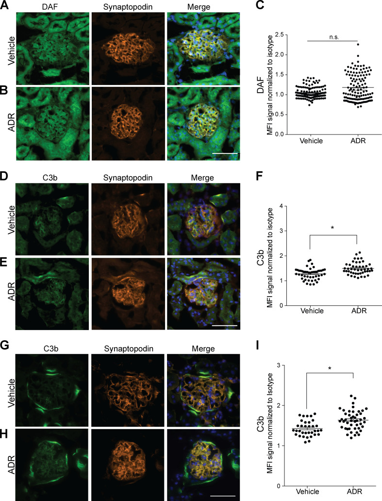
Figure S1.
ADR injection associates with glomerular C3b deposition. (A–F) Representative pictures of glomerular DAF (A and B) and C3b staining (D and E) and data quantification (C and F) of male B6 WT mice at 2 wk after treatment with vehicle or ADR (20 mg/kg, i.v., n = 4). (G–I) Representative glomerular C3b deposition (G and H) and data quantification (I) in kidneys from B6 DAF−/− mice at 2 wk after ADR or vehicle injection (n = 4). All images within the same experiment, including vehicle and ADR, were captured at the same exposure time. n.s., not significant. Scale bars: 50 µm. *P < 0.05. Error bars are SEM.

