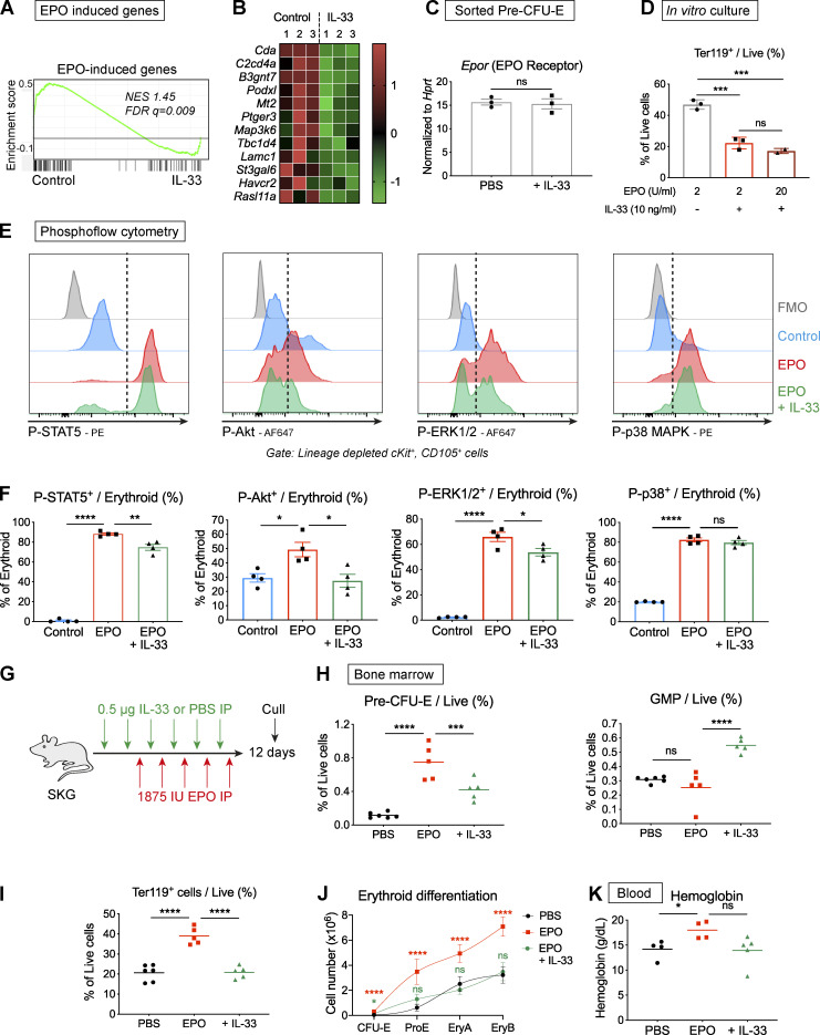
Figure 7.
IL-33 interferes with signal events downstream of the EPO receptor, curtailing EPO-accelerated erythropoiesis. (A) Enrichment plot of GSEA in pre–CFU-Es using a set of genes up-regulated by EPO in CEPs. FDR q, false discovery rate q value; NES, normalized enrichment score. (B) Heatmap showing differential expression of selected genes in pre–CFU-Es with exposure to IL-33 that were induced by EPO in CEP, expressed as row-normalized z scores. Columns show paired replicates from n = three independent experiments, each with n = six mice. (C) Expression of Epor by RT-qPCR in pre–CFU-Es sorted by FACS from littermate SKG mice injected with PBS or IL-33 for 1 wk. Expression normalized to Hprt (mean and SEM of n = three independent experiments, n = two per group, unpaired t test). (D) Proportion of cells expressing Ter119 after culture of FACS-sorted pre–CFU-Es with EPO at 2 U/ml or 20 U/ml, ±IL-33 (mean and SD of technical triplicates, one-way ANOVA with Tukey’s test, representative of two independent experiments). (E) Representative images showing flow cytometric expression of indicated phosphorylated proteins in erythroid progenitors exposed to medium or IL-33 for 6 h then stimulated with EPO. Dashed lines indicate cutoff for positive cells. Representative of four independent experiments. (F) Proportion of erythroid progenitors expressing indicated phosphorylated proteins (mean of n = four independent experiments, n = three replicates per group for each experiment, with mean and SEM, one-way ANOVA with Tukey’s test). (G) Schematic diagram showing injection of littermate SKG mice with IL-33 or PBS, followed by EPO, according to indicated schedule before culling to generate data shown in H–K. (H) Frequencies of indicated progenitor populations in BM (points show individual mice with mean, one-way ANOVA with Tukey’s test, representative of two independent experiments). (I) Frequencies of Ter119+ erythroid cells in BM (points show individual mice with mean, one-way ANOVA with Tukey’s test, representative of two independent experiments). (J) Erythroid differentiation pathways (mean and SD of n = five mice per group, locally weighted scatterplot smoothing (LOWESS) spline, one-way ANOVA with groups compared with PBS group using Tukey’s test, representative of two independent experiments). (K) Blood Hgb concentration (points show individual mice with mean, one-way ANOVA with Tukey’s test, representative of two independent experiments). *, P < 0.05; **, P < 0.01; ***, P < 0.001; ****, P < 0.0001.

