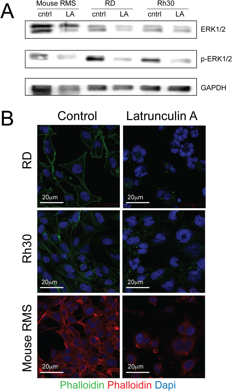
Fig 4. Protein expression levels of ERK and pERK in human and mouse rhabdomyosarcoma cell lines after treatment with Latrunculin A.
Human RD sarcoma cells were exposed to 250nM latrunculin A, human Rh30 RMS cells to 100nM latrunculin A and mouse RMS sarcoma cells to 100nM latrunculin A. Different concentrations were chosen due to differences in latrunculin A sensitivity between sarcoma cell lines. (A) Evaluation of ERK1/2 (42/ 44kDa), p-ERK1/2 (22/ 44 kDa) and GAPDH (37 kDa) by Western blotting demonstrated reduced ERK1/2 phosphorylation in latrunculin A-treated sarcoma cells. (B) The F-actin cytoskeleton was evaluated using phalloidin staining. Actin organization was profoundly disrupted in latrunculin A-treated (top right panel)) vs. control (top left panel) RD cells, latrunculin A-treated (middle right panel) vs. control (middle left panel) Rh30 cells and latrunculin A-treated (bottom right panel) vs. control (bottom left panel) mouse RMS cells. Please also see S1 Raw images.

