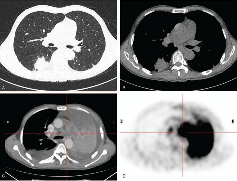
Figure 4.

Male, 59 years old, repeated chest pain in a month and a massive opacity in the right lung for 3 weeks. CT shows a massive opacity with lobulation and speculation in the margin of the lesion, which has a uniform density and mild-moderate enhancement in the superior segment of the right lower lung (A and B). Four months later, the left lung shows large atelectasis with bilateral pleural effusion (C). PET/CT shows atelectasis in the left lung with a high concentration of radioactivity in the maximum SUV value of 19.9 (D).
