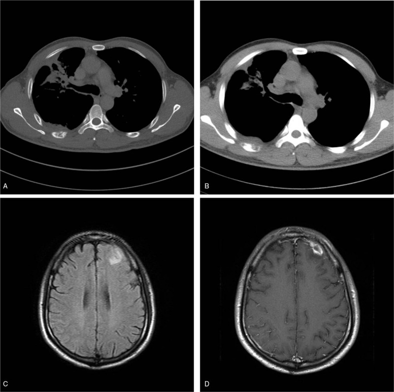
Figure 5.

Male, 37 years old, recurrent cough and fever for 1 month. The new lesions appear in the right upper lung in massive opacities and cord-like shadows with bone destruction (A) and soft tissue mass (B) in the right sixth rib. MRI shows a patchy high signal in the left frontal lobe on T2WI/FLAIR (C). The lesion shows an annular enhancement (D).
