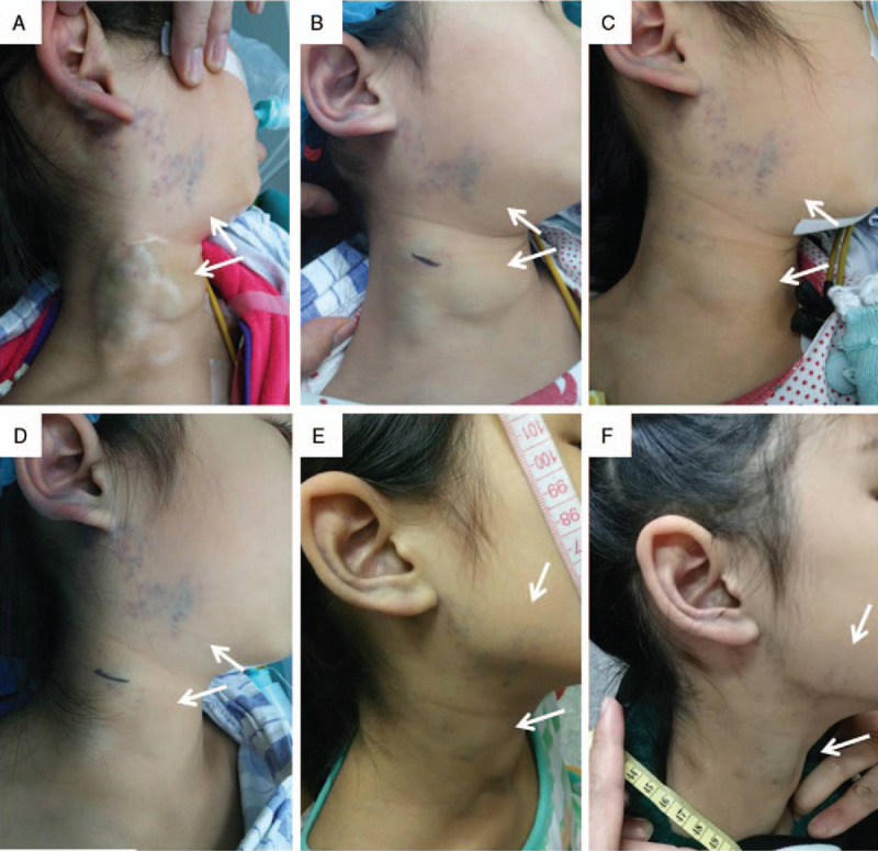
Figure 1.

A 6-year-old female with a large diffuse VM involving the right face and neck region. (A) Lateral view of VM during the first PS session. The lesion was superficial, blue, compressible and soft on palpation. The boundary of the lesion was not clearly defined. (B) One month after the first PS session, during the second PS session. The volume of the lesion did not significantly change. (C) One month after the second PS session, during the third PS session. The lesion was smaller and flat. (D) One month after the third PS session, during the fourth PS session. (E) Three weeks after the fourth PS session, significant reduction in size and color fading of the lesion were noticed (arrow). (F) 18 months after the fourth PS session, although partial VM remained, the desired goal of treatment was achieved. PS = percutaneous sclerotherapy, VM = venous malformation.
