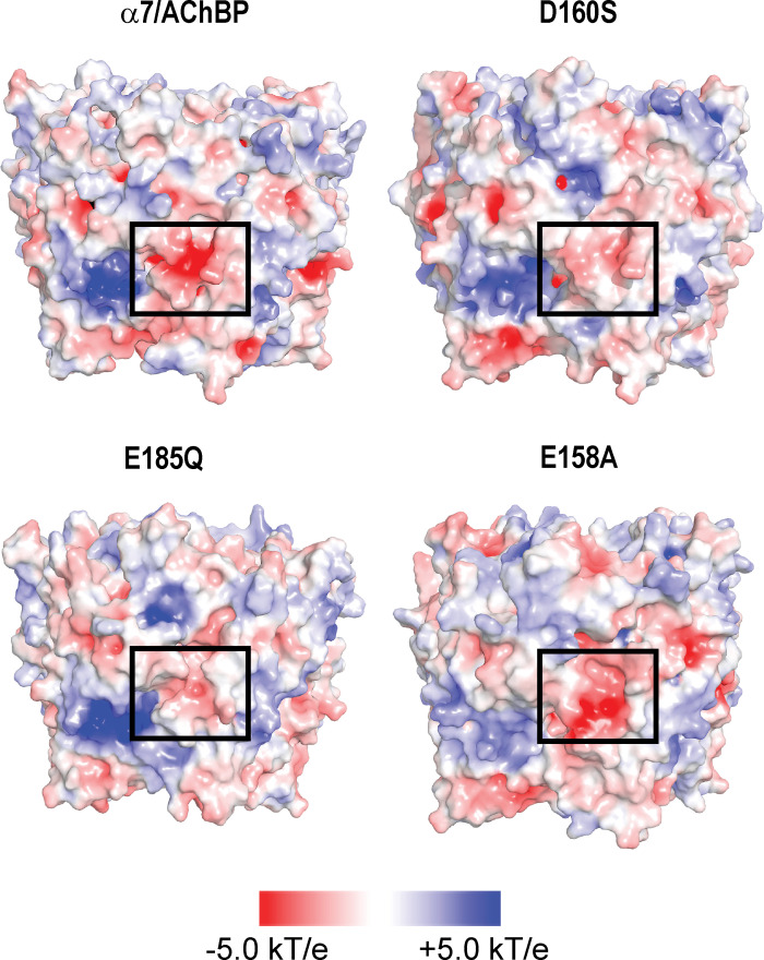
Figure 5.
Electrostatic analysis of wild-type and mutant α7/AChBP pentamers in the absence of calcium. Each structure shows the electrostatic surface potential, computed by solving the Poisson-Boltzman equation, for the structure obtained following a 100-ns MD simulation. Boxed regions encompass residues observed to coordinate Ca2+ in Fig. 4. Red patches represent negative electrostatic potential, blue patches represent positive potential, and white patches represent neutral potential. The mutations D160S and E185Q reduce the negative electrostatic surface potential around the calcium binding site, whereas the E158A mutation maintains a negative electrostatic surface potential at the site. kT/e, electrostatic potential.

