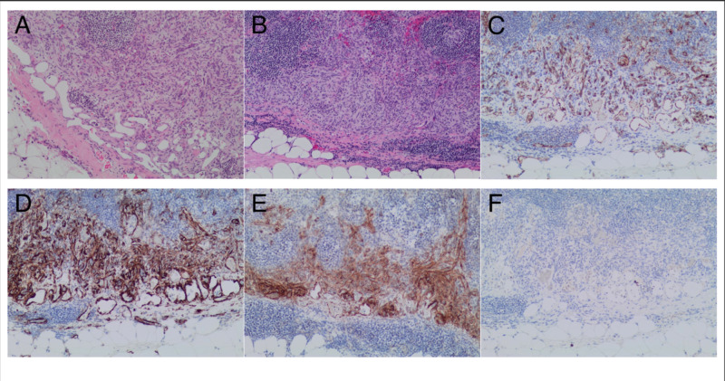
Figure 1. A lymph node involved by a predominantly subcapsular/sinusoidal proliferation of bland spindle cells that are associated with fenestrated capillary proliferation (A, B). The flattened endothelial cell lining is highlighted by CD31 immunostaining, while the spindle cell proliferation is negative for CD31 (C). The spindle cell proliferation shows expression of both SMA and DOG1 (D, E). Both the spindle cell proliferation and the endothelial cells in the capillary proliferation are negative for HHV8 by immunohistochemistry (F).
DOG1, discovered on GIST1; HHV8, human herpesvirus 8; SMA, smooth muscle actin.

