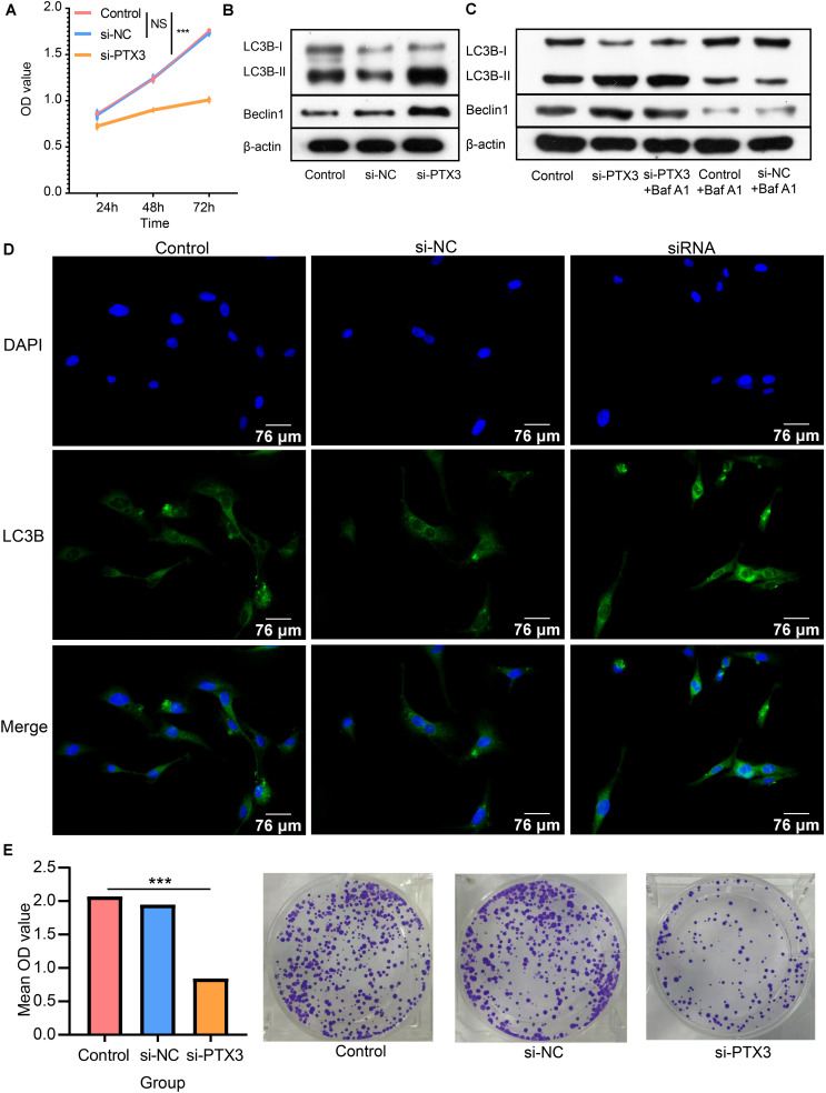
FIGURE 4.
PTX3 affects cells viability and negatively modulate cell autophagy. (A) Cells proliferation ability is evaluated by CCK8 assay at 24 h, 48 h, and 72 h after interfering the expression of PTX3. (B) Western bolt illustrated autophagy related proteins, beclin1 and LC3B, are upregulated when PTX3 expression is inhibited. (C) The autophagy flux assay suggests the transition from LC3B-I to LC3B-II is inhibited when silenced PTX3 expression. (D) U87 MG cells are stained with DAPI (blue) and LC3B (green). Images are captured by laser confocal microscope (×400). (E) The colony-forming assay supports cells viability is inhibited by silencing PTX3 expression. NS, no significantly statistical; *P < 0.05; **P < 0.01; ***P < 0.001.

