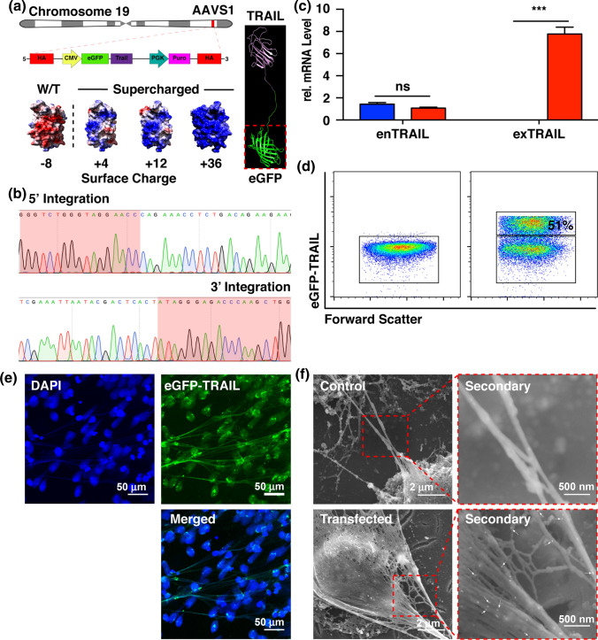
Figure 2.
Engineered neutrophils express supercharged eGFP-TRAIL on NETs during NETosis. (a) Schematic of the insertion site, AAVS1, on chromosome 19. Cartoon representation of eGFP-TRAIL chimeric protein. Surface charge of eGFP ranging from − 4 to + 36 (Red—negative charge, Blue—positive charge. (b) DNA sequencing result of genomic DNA isolated from cells positive for eGFP-TRAIL. (c) relative mRNA level of endogenous (enTRAIL) and exogenous (exTRAIL) TRAIL levels. (d) Flow cytometry of cells expressing eGFP-TRAIL 24 h after nucleofection. (e) Confocal images of NETs decorated with eGFP-TRAIL (Blue—DAPI stain, Green—eGFP-TRAIL). (f) Immuno-gold SEM images of neutrophils expressing eGFP-decorated NETs (White arrows—eGFP-TRAIL).

