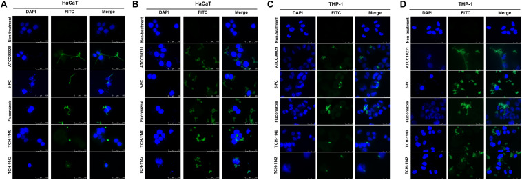
FIGURE 3.
The confocal microscopic images of C. albicans (green) in the host cells (blue) after treatment of 5-FC, fluconazole, and naphthofuranquinones: (A) ATCC90029-infected keratinocytes; (B) ATCC10231-infected keratinocytes; (C) ATCC90029-infected macrophages; and (D) ATCC10231-infected macrophages.

