Figure 4.
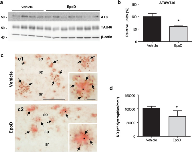
Tau pathology decreased in the hippocampus of EpoD-treated mice. (a,b) Representative western blot and quantitative analysis of the phospho-tau ratio (AT8/TAU46) from APP/PS1Veh (n = 4) and APP/PS1EpoD (n = 7) mice. β-Actin was used as the loading control. For quantification, ratio was referred to vehicle group. The ratio of phospho-tau/total tau was significantly reduced in treated mice (t test, *p < 0.05). Uncropped blots are shown in Supplementary Fig. S7. (c) AT8-positive dystrophies (arrows) were located surrounding Aβ plaques (Congo red, asterisk). Quantitative analysis (d) showed a significant decrease in the numerical density (ND) of AT8-positive dystrophic neurites (t test, *p < 0.05) in CA1 region from EpoD (c2) compared to vehicle mice (c1). so stratum oriens, sp stratum pyramidale, sr stratum radiatum. Scale bars: (c1) and (c2) 200 μm; insets 20 μm.

