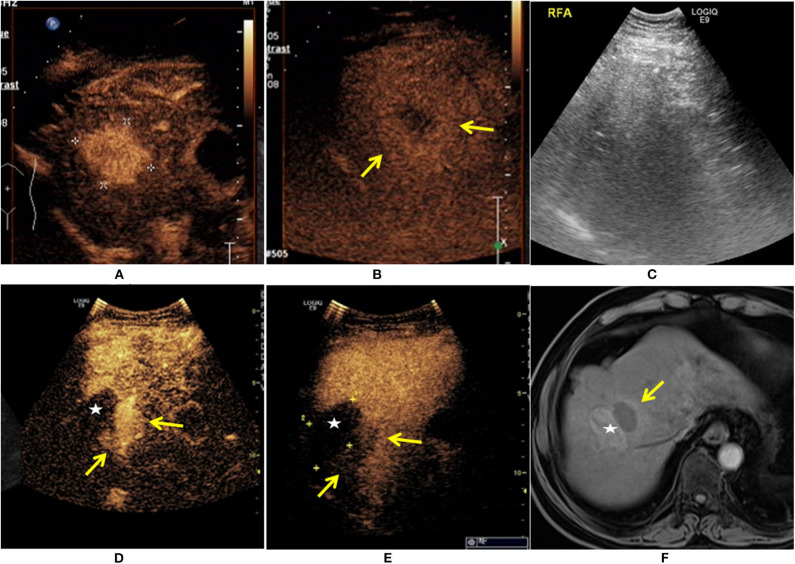
Figure 1.
A 65-year-old man was diagnosed with HCC. The patient had a 30-year history of hepatitis B. The contrast-enhanced ultrasound (CEUS) showed a single lesion measuring 4.1 × 3.6 cm in size (+) enhanced at arterial phase (A), and the mild washout in lesion was found at the late phase ( ) (B). The patient received ultrasound-guided radiofrequency ablation (RFA) treatment (C). Eleven months after RFA, there was an irregular enhancement (
 ) around the ablation zone (★), and marked washout at the late phase (
 ) (D,E). The local recurrence after RFA was also demonstrated at enhanced MRI (
 ) (F).

