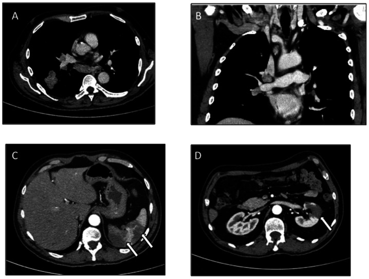
Figure 1.
Baseline CT scan. (A and B) Thoracic CT scan showed a mass in the lower lobe of the right lung, with ipsilateral ilo-mediastinal (subcarinal) and right infra-clavear pathological lymph nodes. (C and D) Abdominal CT scan documented signs of previous, non-datable multiple splenic and renal infarctions (white arrows), likely attributable to an acute/subacute phase of infarction.

