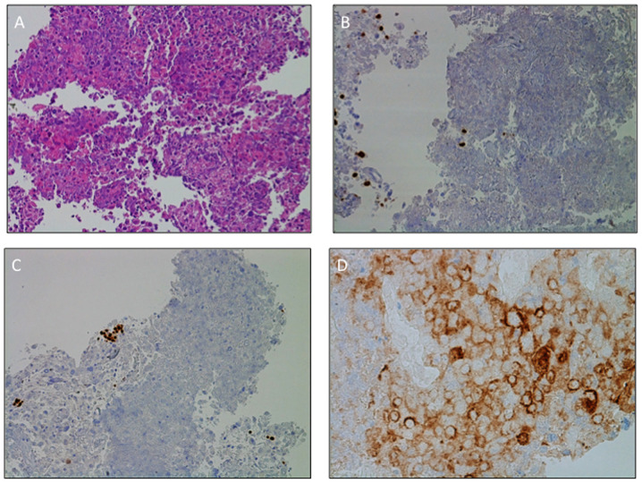
Figure 3.
Morphological and immunohistochemistry diagnosis of large-cell carcinoma of the lung. (A) Hematoxylin and eosin staining documenting malignant large pleomorphic cells with a moderate to abundant amount of cytoplasm, vesicular nuclei, prominent nucleoli and necrosis. IHC staining of (B) TTF1 and (C) p40 revealed expression of the two markers on pneumocytes (TTF1) and basal cell (p40) only, thus supporting the diagnosis of large-cell carcinoma of the lung. The negativity of TTF1 and p40 precluded the diagnosis of lung adenocarcinoma or squamous cell carcinoma, respectively. Tumors lacked morphological and immunohistochemical evidence of glandular, squamous or neuroendocrine differentiation. (D) Programmed death-ligand 1 expression levels on the surface membrane of 55% of tumor cells. Magnification, (A-C) ×20 and (D) ×40. TTF1, transcription termination factor 1.

