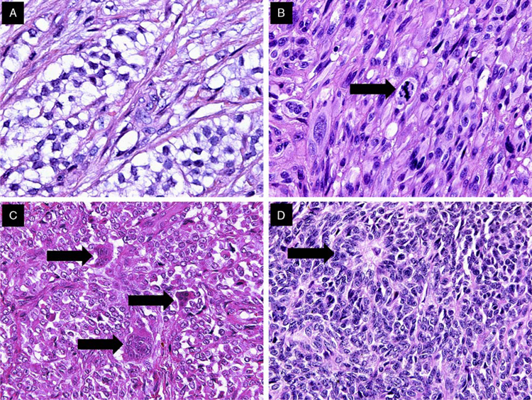
FIGURE 4.
Examples of histologic features of GNETs. A, Focal clearing of the cytoplasm was relatively uncommon. B, Admixed spindle and epithelioid tumor cells; mitosis (arrow). C, Multinucleated osteoclast-like giant cells (arrows), were observed in half of the cases. D, Occasional rosette-like structures (arrow) were identified in several cases.

