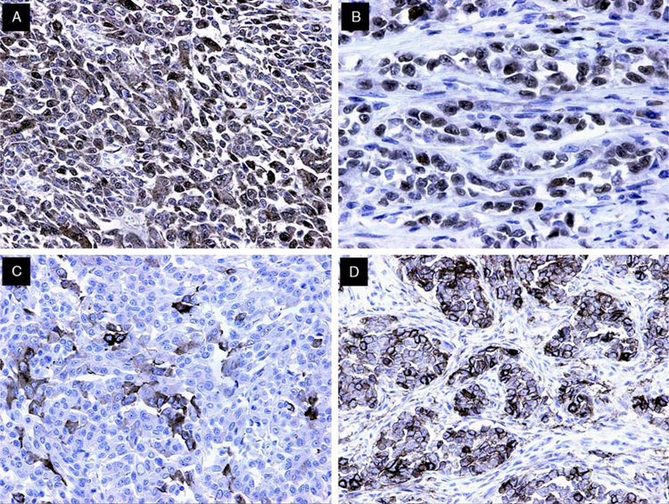
FIGURE 5.
Immunohistochemical findings in GNET. A, S-100 protein expression was mainly strong and diffuse, in a nuclear and cytoplasmic distribution. B, SOX10 staining was uniformly strongly positive in the majority of tumor cells. C, Synaptophysin expression was common, with most cases displaying focal but strong cytoplasmic staining. D, CD56 expression was variable. Note staining of neoplastic cells arranged in nests surrounded by desmoplastic stroma.

