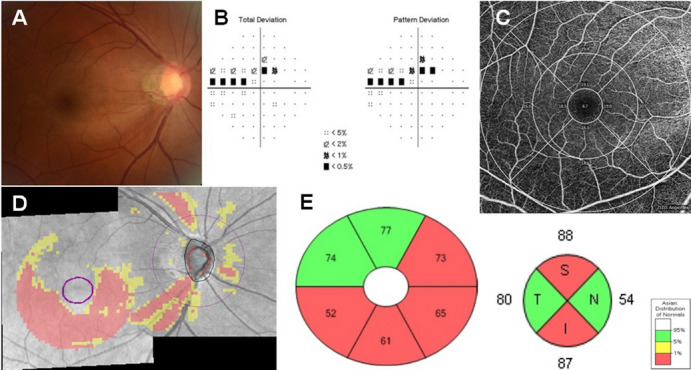
Figure 1.

Inferior disc excavation with inferior nerve fibre layer wedge defect of right eye of a 52-year-old woman with POAG (a). Visual field results revealed defects at the corresponding superonasal site (b). OCT angiography image (Cirrus HD-OCT , version 10.0.0.14618) of a 6 × 6 mm scan exhibited the superficial vessels at the macula. The grids represented the sectors across which the vessel length densities (/mm) (c) were calculated. The area between the inner two circles in the 6 × 6 mm scan represented the inner sectors, and the area between the outer two circles represented the outer sectors. Combined RNFL and GCIPL deviation maps indicated structural glaucomatous damage (d). OCT scan showed the GCIPL thickness (μm) provided by the ganglion cell analysis report (e, left) and RNFL thickness (μm) (e, right) in each sector. GCIPL ganglion cell-inner plexiform layer; OCT optical coherence tomography. I inferior; IN inferonasal; IT inferotemporal; N nasal; S superior; SN superonasal; ST superotemporal; T temporal.
