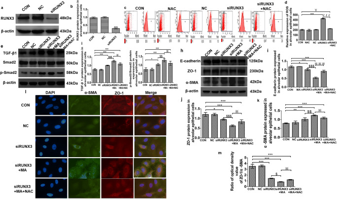
Fig. 5.
RUNX3-related oxidative EMT induced by MA through TGF-β signaling. a, b The expression of RUNX3 after transfection by Western blot assay. c, d ROS levels in the alveolar epithelial cells by flow cytometry. The mean value in P1 is the average fluorescence intensity, as a statistical indicator, representing the expression of ROS. (e) TGF-β signaling expression by Western blot. f TGF-β 1 expression in alveolar epithelial cells in the various groups. g p-Smad2/Smad2 expression in alveolar epithelial cells in the various groups. h–k EMT marker proteins E-cadherin, ZO-1, and α-SMA expressed in alveolar epithelial cells in the various groups. l, m Immunofluorescence assay of α-SMA and ZO-1 in alveolar epithelial cells. After transfection for 72 h, the cells were treated with MA (5 mM, 48 h) and/or NAC (5 mM, 1 h), respectively. Data are presented as the mean ± SD. *P < 0.05, **P < 0.01, ***P < 0.001 vs. CON group; #P < 0.05 vs. NAC; $P < 0.05, $$P < 0.01, $$$P < 0.001 vs. siRUNX3; ※P < 0.05, ※※※P < 0.001 vs. siRUNX3+MA. CON, control; NC, negative control (empty plasmid); siRUNX3, siRNA against RUNX3; NAC, N-acetyl cysteine; MA, methamphetamine

