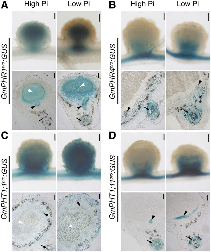
Figure 2.
Tissue-specific expression of GmPHR-GmPHT1 modules in soybean nodules. GmPHR1 (A), GmPHR4 (B), GmPHT1;1 (C), and GmPHT1;11 (D) promoters were individually fused to a GUS reporter gene, and the resulting vectors were transferred into soybean hairy roots that were cultured under high (left images) or low (right images) Pi concentrations. Nodules were stained with X-Gluc, and whole nodule tissue (top images) or semithin sections (bottom images) were observed with a microscope. Photographs were taken from sections of different nodules produced from at least two independent biological experiments. A single representative result is shown. Black arrows indicate nodule vascular bundles, white arrowheads indicate nodule infected tissues, and black arrowheads indicate root vascular tissues. Bars = 200 μm (top images) and 100 μm (bottom images).

