Figure 7.
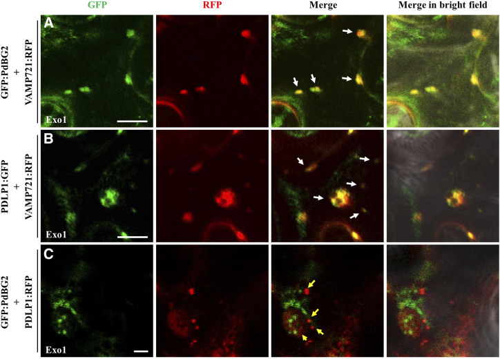
PdBG2 and PDLP1 proteins are secreted with different cargo machinery. A, Confocal images show colocalization between GFP:PdBG2 and VAMP721:RFP after Exo1 (10 μm) treatment (12 h). B, Confocal images show colocalization between PDLP1:GFP and VAMP721:RFP after Exo1 (10 μm) treatment (12 h). In A and B, white arrows indicate that GFP and RFP signals were perfectly merged. C, Confocal images show different localization between GFP:PdBG2 and PDLP1:RFP after Exo1 (10 μm) treatment (12 h). Yellow arrows indicate that GFP and RFP signals were not colocalized. The fusion proteins were transiently expressed in N. benthamiana leaf epidermal cells. Bars = 10 µm (A and B) and 2 µm (C)

