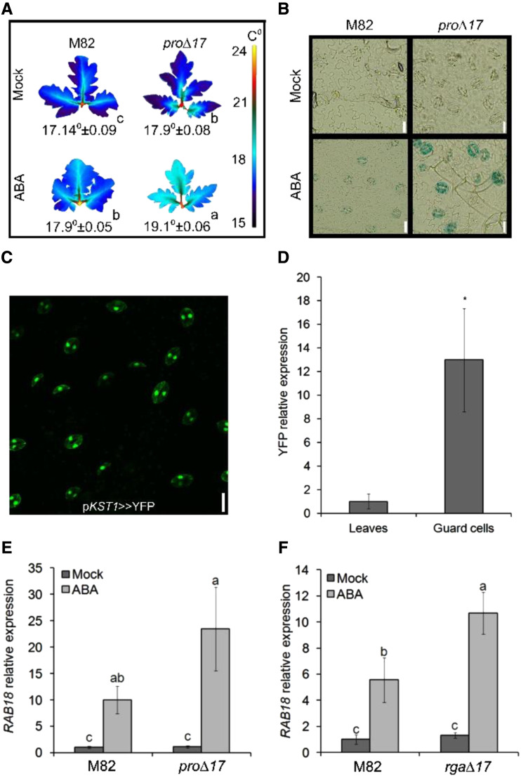
Figure 1.
PRO promotes ABA responses in guard cells. A, Thermal imaging of leaves (leaf no. 4 below the apex) taken from M82 and 35S:pro∆17 treated or not (Mock) with 10 μm of ABA. Leaves were digitally extracted for comparison. Number below leaves are the calculated leaf-surface temperature and the values are means of three plants, measured 20 times ± se. Small letters above the numbers represent significant differences between respective treatments (Tukey–Kramer HSD test, P < 0.05). B, Representative images of GUS staining of epidermal peels treated or not (Mock) with 10 μm of ABA. Peels were taken from leaf no. 4 below the apex of M82 and 35S:pro∆17 expressing the reporter GUS under the regulation of the MAPKKK18 promoter. C, YFP signal in guard cells of pKST1>>YFP transactivated epidermal peel. Scale bars = 20 μm. D, YFP expression in whole leaf tissue and guard-cell–enriched samples. Values are means of four biological replicates ± se. Stars above the columns represents significant differences between respective treatments by Student’s t test (P < 0.05). E and F, RT-qPCR analysis of RAB18 expression in guard-cell–enriched samples isolated from leaves no. 3 and 4 below the apex of M82 and 35S:pro∆17 (E) or 35S:rga∆17 (F). Values in E and F are means of four biological replicates ± se. Small letters above the columns represent significant differences between respective treatments by Tukey–Kramer HSD (P < 0.05). The value for leaves in D was set to 1 and the value for M82 Mock in E and F was set to 1.

