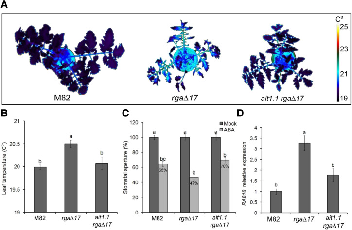
Figure 5.
ait1.1 suppressed the effect of PRO on ABA responses in guard cells. A, Thermal imaging of M82, 35S:rgaΔ17 (rgaΔ17), and rgaΔ17 ait1.1. Images were digitally extracted for comparison. B, Leaf-surface (leaves no. 3 and 4 below the apex) temperature of M82, rgaΔ17, and rgaΔ17 ait1.1. plants. Values are means of three replicates measured 20 times ± se. C, Stomatal aperture in M82, rgaΔ17, and rgaΔ17 ait1.1 epidermal peels (from leaves no. 3 and 4 below the apex) treated or not treated (Mock) with 10 μm of ABA. One hour after the ABA treatment, stomatal aperture was measured. Values are mean percentage of Mock of four replicates, each with ∼100 measurements (stomata) ± se. D, RT-qPCR analysis of RAB18 expression in M82, rgaΔ17, and rgaΔ17 ait1.1 guard cells, isolated from leaves no. 3 and 4 below the apex. Values are means of four biological replicates ± se. Different letters above the columns in B and D represent significant differences between lines by Student’s t test (P < 0.05). Different letters above the columns in C represent significant differences between lines and treatments by Tukey–Kramer HSD test (P < 0.05). The values for M82 were set to 1.

