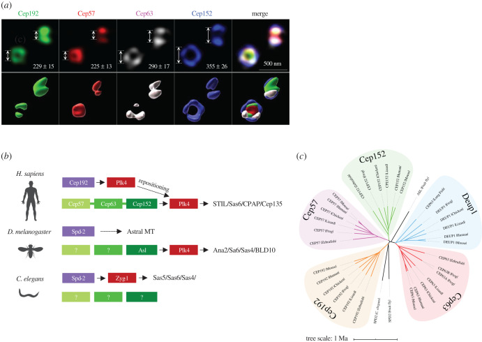
Figure 1.
Organizational features of inner PCM scaffolds critical for the procentriole assembly pathway. (a) Three-dimensional structured illumination microscopy (3D SIM) analysis showing the pericentriolar localization patterns of Cep192, Cep57, Cep63 and Cep152 in U2OS cells. Cells fixed with 4% paraformaldehyde were coimmunostained with antibodies against Cep192 N-terminal, Cep57 C-terminal, Cep63 full-length and Cep152 N-terminal regions. Average diameters quantified from greater than 40 images are shown. 3D surface-rendering was carried out with Imaris v.8.4.1 (Bitplane). (b) Schematic diagram showing upstream scaffolds critical for recruiting Plk4/Zyg-1 in humans, flies and worms. Components are positioned relative to their locations from the centriolar axis. In Homo sapiens, Plk4 initially recruited to the inner Cep192 scaffold relocalizes to the outer Cep152 (repositioning) before inducing downstream events. The Cep57-Cep63-Cep152 complex is linked with a thick line. Question marks indicate no apparent orthologues found. (c) A phylogenetic tree for inner PCM scaffolds, including the Cep63 paralogue, Deup1, shown to be critical for deuterosome-dependent centriole amplification in multiciliated cells [31]. Note that vertebrate Cep152 and Cep192 greatly diverge from D. melanogaster Asl and C. elegans Spd-2, respectively.

