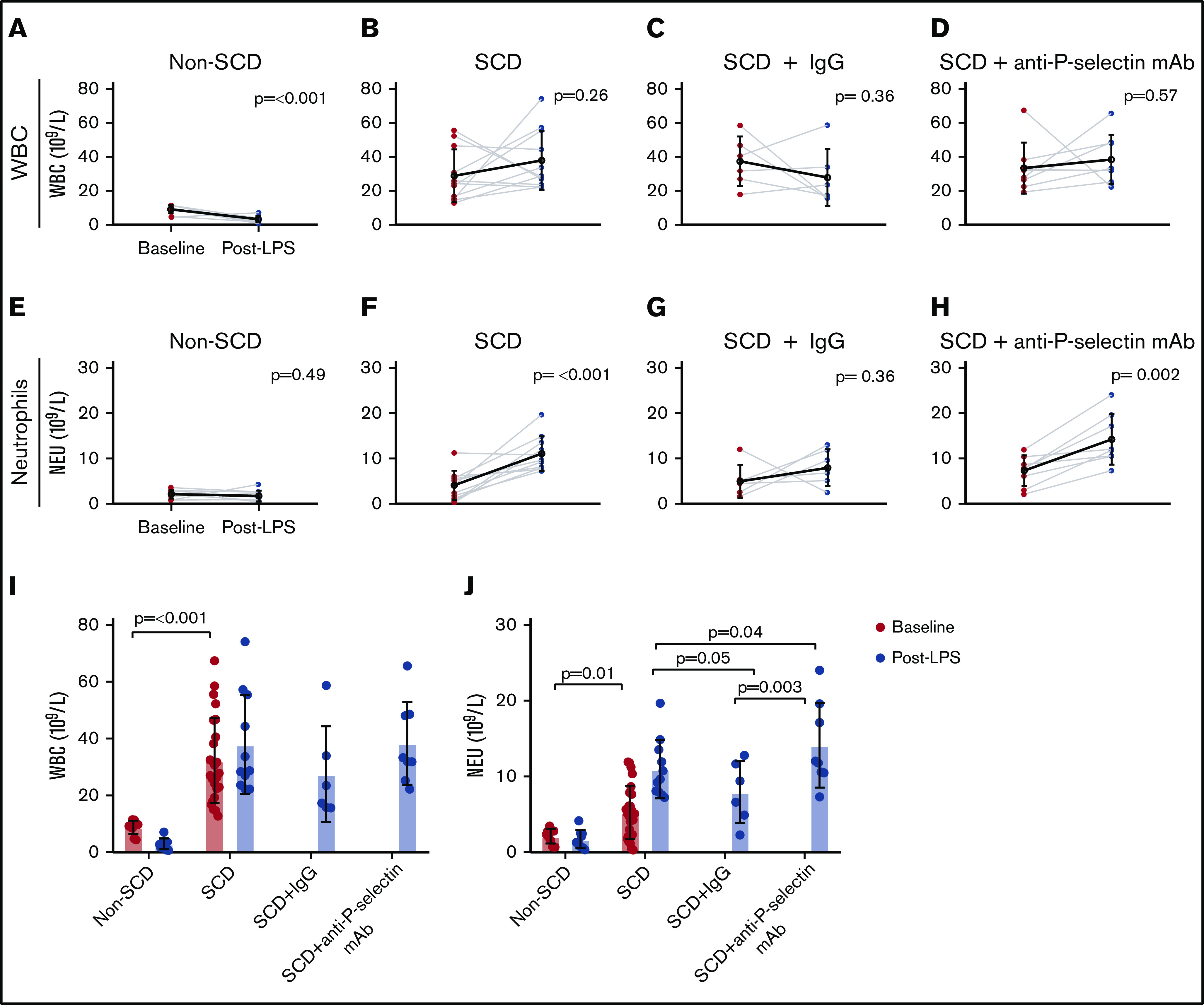
Figure 4.

WBC and neutrophil counts for the non-SCD (control), SCD, and SCD mice treated with either IgG isotype control or anti-P-selectin mAb. (A-D) WBC counts (× 109/L) at baseline and post-LPS challenge for non-SCD (A), SCD (B), SCD + IgG isotype control (C), and SCD + anti-P-selectin mAb (D). (E-H) Neutrophil (NEU) counts (× 109/L) at baseline and post-LPS for non-SCD (E), SCD (F), SCD + IgG isotype control (G), and SCD + anti-P-selectin mAb (H). (A-H) Baseline and post-LPS means are shown as a black hollow circle with SD error bars. Baseline and post-LPS groups were compared using a paired 2-tailed Student t test. P < .05 was considered statistically significant. (I) Baseline and post-LPS WBC (× 109/L) measurements for all conditions. Non-depicted P values: post-LPS non-SCD was significantly different from post-LPS SCD (P ≤ .001), SCD + IgG (P = .002), and SCD + anti-P-selectin mAb (P ≤ .001). (J) Baseline and post-LPS neutrophil (× 109/L) measurements for all conditions. Non-depicted P values: post-LPS non-SCD was significantly different from post-LPS SCD (P ≤ .001), SCD + IgG (P = .002), and SCD + anti-P-selectin mAb (P ≤ .001). (I-J) Bars represent mean ± SD. Baseline analysis between SCD and non-SCD used a 2-tailed Student t test. One-way ANOVA with multiple comparisons by controlling FDR by a 2-stage linear step-up procedure of Benjamini, Krieger, and Yekutieli was used for the post-LPS analysis. FDR-corrected P < .05 was considered statistically significant.
