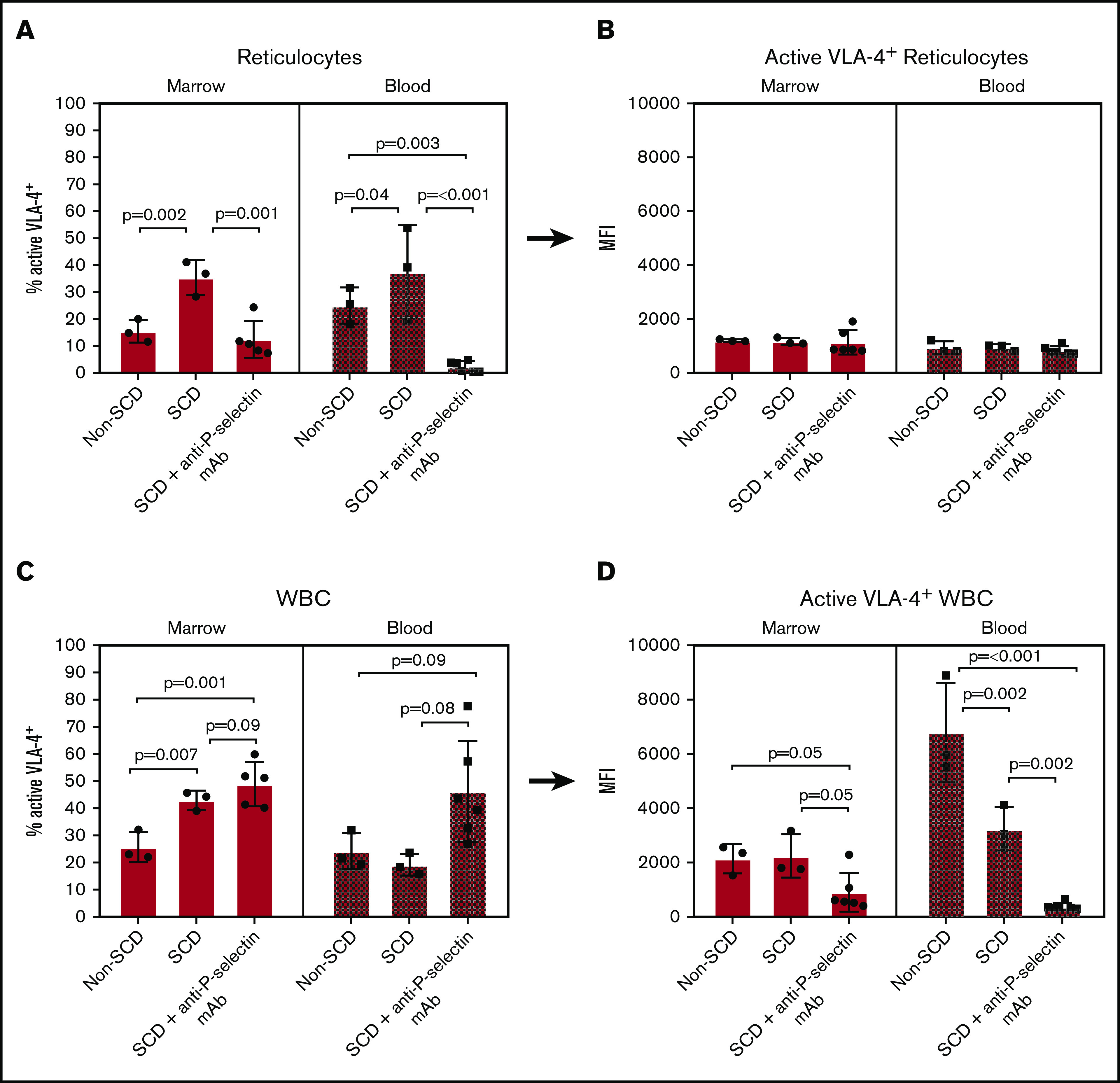
Figure 5.

Flow cytometry of isolated bone marrow and blood cells labeled with Cy3-LLP2A post-LPS challenge. (A-B) Reticulocyte-gated measurements. (A) Percent of reticulocytes that express active VLA-4 in the bone marrow and blood. (B) The MFI of Cy3-LLP2A-bound VLA-4 on VLA-4+ reticulocytes. (C-D) WBC-gated measurements. (C) Percent of WBCs that express active VLA-4 in the bone marrow and blood. (D) The MFI of Cy3-LLP2A-bound VLA-4 on VLA-4+ reticulocytes. (A-D) Bars represent mean ± SD. Data were analyzed by 1-way ANOVA with multiple comparisons by controlling FDR by a 2-stage linear step-up procedure of Benjamini, Krieger, and Yekutieli. FDR-corrected P < .05 was considered statistically significant.
