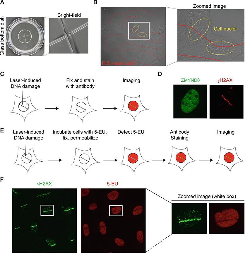
Fig. 2.
Immunofluorescence analysis of endogenous protein recruitment and transcription in the DNA damage site. (a) Experimental setup for laser microirradiation of fixed samples. Left: etched crossed line on glass bottom dish. Right: zoomed image using 60× oil immersion objective and bright-field microscopy. (b) Free drawn ROI line on U2OS human cancer cells within glass bottomed dishes viewed by bright-field microscopy. Red line indicates the ROI and yellow circle indicates the cell nuclei. (c) Schematic illustration for analysis of endogenous protein recruitment and transcription in DNA damage site. (d) Endogenous ZMYND8 translocation to DNA damage site. DNA damage was induced by laser microirradiation and stain with ZMYND8 and γH2AX antibodies. (e) Schematic of nascent transcription and laser microirradiation technique. (f) Transcription analysis in the DNA damage site using 5-EU staining. γH2AX is marker for the DNA damage region

