Figure 1.
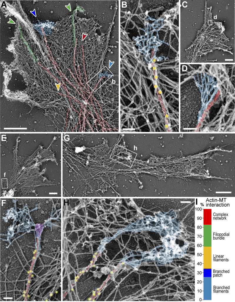
Branched actin networks in growth cones can be physically linked to MT tips. (A–H) PREM of growth cones of DIV1 (C and D) or DIV2 (A, B, and E–H) rat hippocampal neurons. Boxed regions in panels A, C, E, and G are enlarged in panels B, D, F, and H, respectively. Color coding: microtubules (red), branched actin networks associated with microtubules (light blue), filopodial bundles (green), α-tubulin immunogold (yellow), and putative +TIPs (purple). (I) Percentage of indicated actin filament arrays associated with microtubule ends in growth cones; n = 87 microtubules in 15 growth cones from six DIV2 neurons labeled with α-tubulin immunogold; one experiment was quantified; six additional independent experiments with DIV2/3 neurons, either unlabeled (four experiments) or labeled by α-tubulin immunogold (two experiments), were assessed qualitatively. Examples of these arrays are shown in A by arrowheads of matching colors. Scale bars, 1 µm (A, C, E, and G); 100 nm (B, D, F, and H).

