Figure 6.
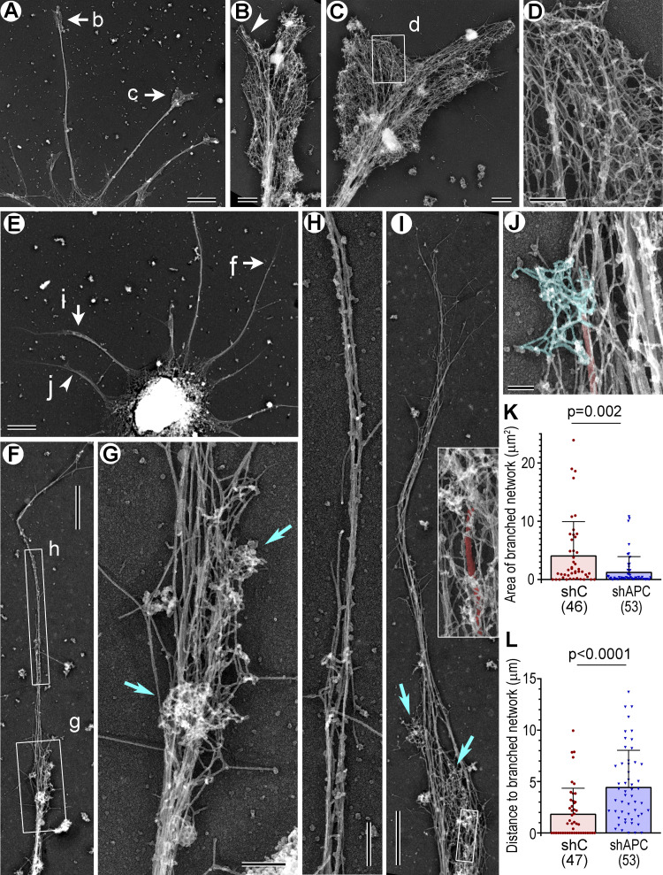
PREM of control and APC-depleted rat hippocampal neurons. Labeled arrows and boxes indicate regions enlarged in respective panels. (A–D) DIV6 neuron treated with control shRNA. (A) A part of the neuron with several dendrites. (B and C) Enlarged growth cones; arrowhead in B indicates a filopodium. (D) Enlarged view of branched actin network. (E–J) DIV6 neuron treated with APC sh1. (E) A part of the neuron with several dendrites. (F and I) Dendrites containing small patches of branched actin (box g in F; cyan arrows in I) and long unbranched extensions at the tips. (G and H) Enlarged views of branched actin patches (G, cyan arrows) and the unbranched actin extension, in which microtubules are not detectable (H). (I) Inset: Enlarged boxed region from the main panel. The most distal microtubule in this dendrite (red) associates with two patches of branched network, at the tip and slightly below. (J) A small region of the dendrite containing a microtubule (red) that terminates in a patch of branched network. (K) Area of most distal regions of cohesive branched network. (L) Lengths of the longest unbranched extensions at neurite tips. Mean ± SD; n is shown for each sample; Mann–Whitney test. One experiment was quantified; two additional experiments were evaluated qualitatively. Scale bars, 5 µm (A and E), 1 µm (F and I), 500 nm (B and C), 200 nm (D, G, and H), and 100 nm (J).

