Figure 2.
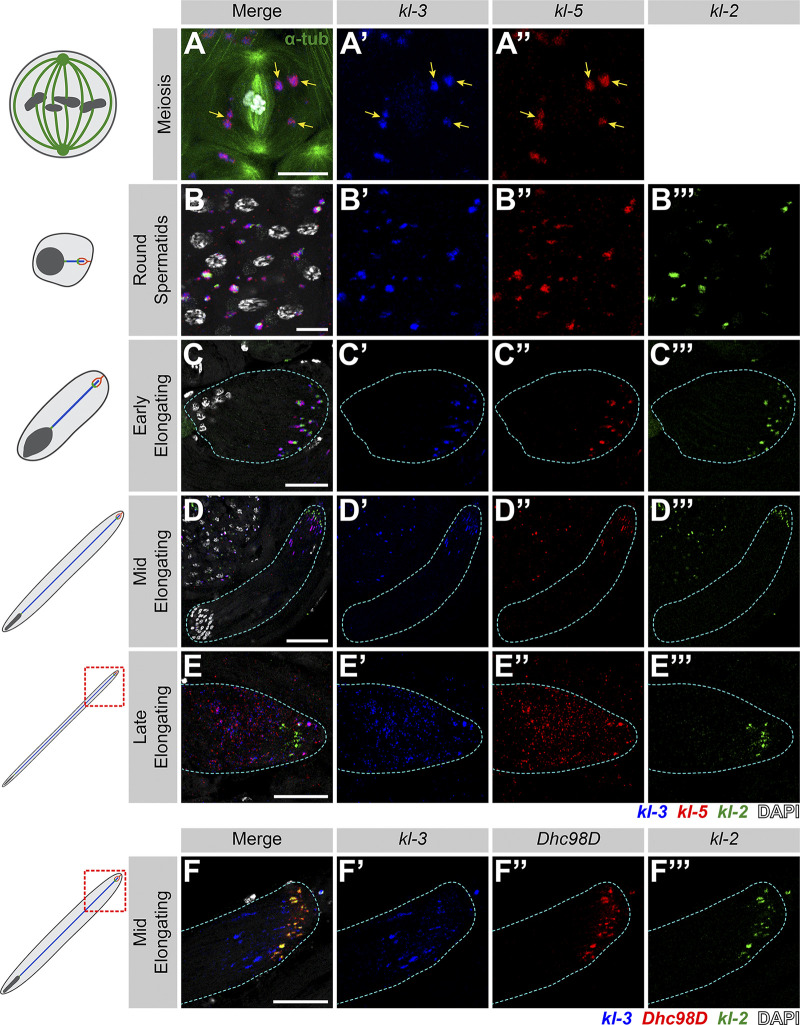
kl-granules segregate during the meiotic divisions and localize to the distal end of elongating spermatids. (A) smFISH against kl-3 and kl-5 during meiosis. Shown are kl-3 (blue), kl-5 (red), α-tubulin-GFP (green), DAPI (white), and kl-granules (yellow arrows). Scale bar: 10 µm. (B–E) smFISH against kl-3, kl-5, and kl-2 during spermiogenesis. The round spermatid (B), early elongating spermatid (C), mid elongating spermatid (D), and late elongating spermatid (E) stages are shown. Shown are kl-3 (blue), kl-5 (red), kl-2 (green), DAPI (white), and a spermatid cyst (cyan dashed line). Scale bars: 10 µm (B), 25 µm (C and E), or 50 µm (D). (F) smFISH against kl-3, Dhc98D, and kl-2 in mid elongating spermatids. kl-3 (blue), Dhc98D (red), kl-2 (green), DAPI (white), spermatid cyst (cyan dashed line). Scale bar: 25 µm.

