Figure 6.
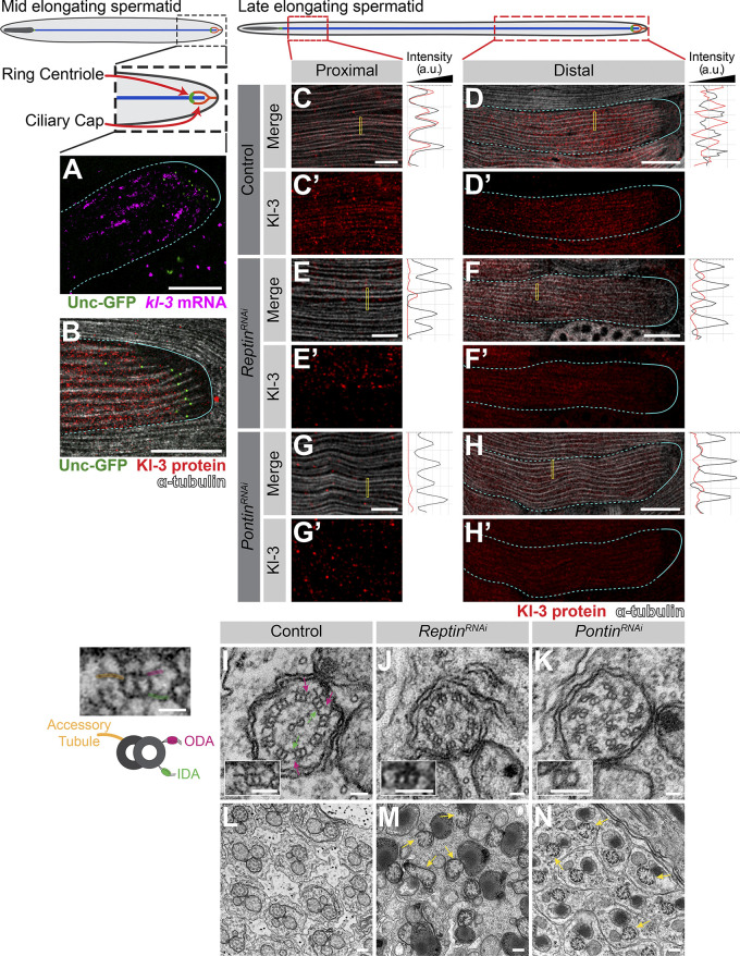
kl-granule formation and localization are required for cytoplasmic cilia maturation. (A) smFISH against kl-3 in flies expressing Unc-GFP. Shown are kl-3 (magenta), Unc-GFP (ring centriole, green), and a spermatid cyst (cyan, dashed line: cytoplasmic region; solid line: compartmentalized region). Scale bar: 20 µm. (B) Kl-3-3X FLAG protein in flies expressing Unc-GFP. Shown are Kl-3 (red), Unc-GFP (ring centriole, green), α-tubulin (white), and spermatid cyst (cyan, dashed line: cytoplasmic region, solid line: compartmentalized region). Scale bar: 20 µm. (C–H) Kl-3-3X FLAG protein expression in control (C and D), rept RNAi (bam-gal4>UAS-reptKK105732; E and F), and pont RNAi (bam-gal4>UAS-pontKK101103; G and H) proximal (C, E, and G) and distal (D, F, and H) regions of late elongating spermatids. Shown are Kl-3 (red), α-tubulin-GFP (white), and a spermatid cyst (cyan, dashed line: cytoplasmic region; solid line: compartmentalized region). Intensity plots are shown for the regions within the yellow rectangles. Scale bars: 5 µm (C, E, and G) or 25 µm (D, F, and H). (I–N) Transmission EM images of control (I and L), rept RNAi (bam-gal4>UAS-reptKK105732; J and M), and pont RNAi (bam-gal4>UAS-pontKK101103; K and N) axonemes. Magenta arrows, ODA; green arrows, IDA; yellow arrows, broken axonemes. The control single doublet enlarged image is duplicated to the left of the figure and colored to match the diagram. Scale bars: 50 nm (I–K), 200 nm (L–N), or 25 nm (diagram left of I).

