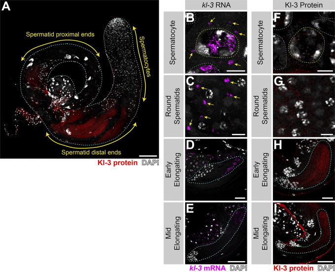
Figure S4.
Kl-3 translation correlates with kl-granule dissociation and is enriched at the distal end. (A) Kl-3 3X FLAG protein expression in a wild-type testis. Shown are Kl-3 (red), DAPI (white), and testis outline (cyan dashed line). Scale bar: 100 µm. (B–E) smFISH for kl-3 in the indicated developmental stages. Shown are kl-3 (magenta), DAPI (white), SC nuclei (yellow dashed line), neighboring SC nuclei (white dashed line), a spermatid cyst (cyan dashed line), and kl-granules (yellow arrows). (F–I) Kl-3 3X FLAG protein in the indicated developmental stages. Shown are Kl-3 (red), DAPI (white), SC nuclei (yellow dashed line), neighboring SC nuclei (white dashed line), and a spermatid cyst (cyan dashed line). Scale bars: 10 µm (B, C, F, and G), 25 µm (D and H), and 50 µm (E and I).

