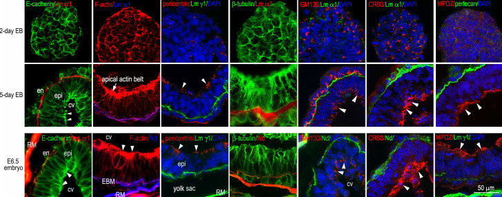
Figure S1.
EB differentiation recapitulates the process of epiblast polarization during mouse peri-implantation development. 2- and 5-d EBs as well as E6.5 mouse embryos were immunostained for E-cadherin, pericentrin, β-tubulin, GM130, CRB3, and MUPP1. The basement membrane was identified by immunofluorescence of nidogen (Nd), perlecan, or the laminin α1 or γ1 chain. F-actin was visualized with rhodamine-phalloidin. The nucleus was counterstained with DAPI. As EBs begin to form the epiblast epithelium, the adherens junction receptor E-cadherin gradually concentrates on the apical side of the epiblast (arrowheads). Cortical F-actin is reorganized to form an apical actin belt (the arrow and arrowheads) and longitudinal filaments. Pericentrin, a component of the microtubule organization center (MTOC), also moves to the apex, where it marks the minus ends of microtubules. Microtubules, labeled with β-tubulin antibody, form a longitudinal array parallel to the lateral plasma membranes. Meanwhile, nuclei are repositioned from the center to the basal side, while the Golgi protein GM130 is localized to the apical side of the epiblast (arrowheads). The polarity markers CRB3 and MPDZ are translocated from a vesicular compartment and the cell cortex, respectively, to the apical surface of the epiblast (arrowheads). cv, central cavity; EBM, embryonic basement membrane; en, endoderm; epi, epiblast; RM, Reichert’s membrane.

