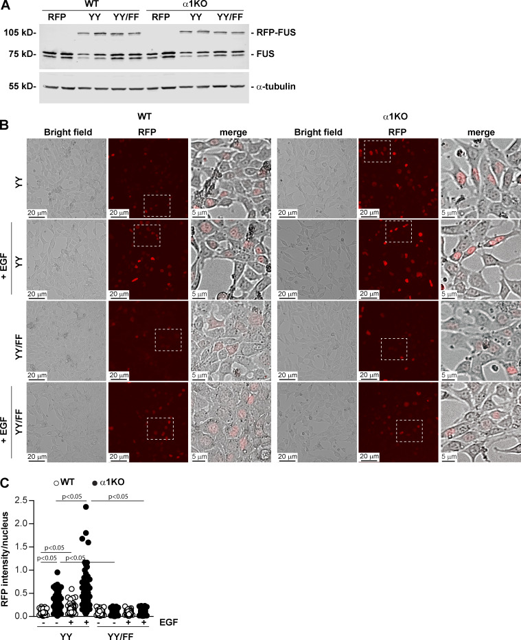
Figure 6.
EGF-mediated FUS phosphorylation induces FUS nuclear translocation. (A) WT and Itgα1KO mesangial cells were transiently transfected with pRFP-C1 empty vector (RFP), or pRFP-C1 vector fused to WT murine FUS (YY) or FUS construct with mutated tyrosines 6 and 296 (YY/FF). After 48 h, cell lysates (20 µg/lane) were analyzed for levels of endogenous and RFP-FUS (two transfections are shown). (B and C) Serum-starved mesangial cells expressing the RFP-FUS constructs described in A were left unstimulated or stimulated with EGF for 30 min (20 ng/ml). Before and after treatment, the cells were imaged, and the nuclear RFP intensity was analyzed using ImageJ. Values in C represent RFP intensity/nuclear area. Circles represent single nuclei (n = 200–208 with two experiments performed) and bars represent mean ± SD. One-way ANOVA and two-tailed t test were used for statistical analysis.

