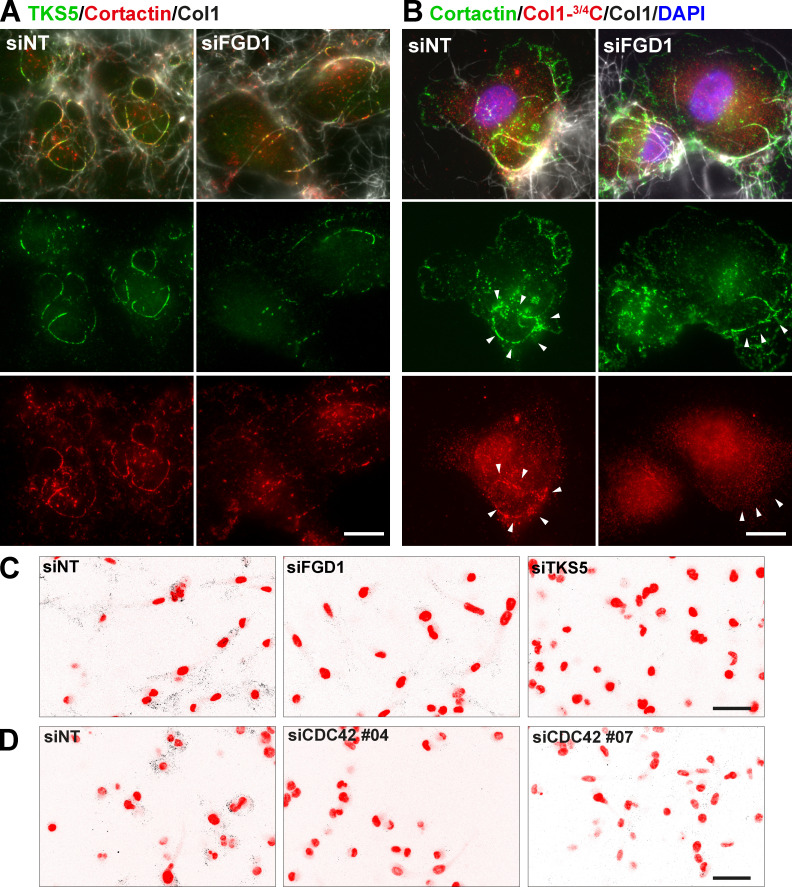
Figure S3.
Pericellular collagen degradation requires the components of the TKS5/FGD1/CDC42 axis. (A) MDA-MB-231 cells treated with FGD1 siRNA were cultured on a fibrillary layer of type I collagen (gray) and stained for TKS5 (green) and cortactin (red). (B) Cells as in B stained for cortactin (red). Cleaved collagen was stained with the Col1-3/4C antibody (red). Cell nuclei were stained with DAPI (blue). Scale bars, 10 µm. (C and D) Representative images of pericellular collagenolysis detected with Col1-3/4C antibody (black signal in the inverted images). Nuclei were stained with DAPI (pseudo-colored in red). Scale bar, 50 µm.

