Figure S5.
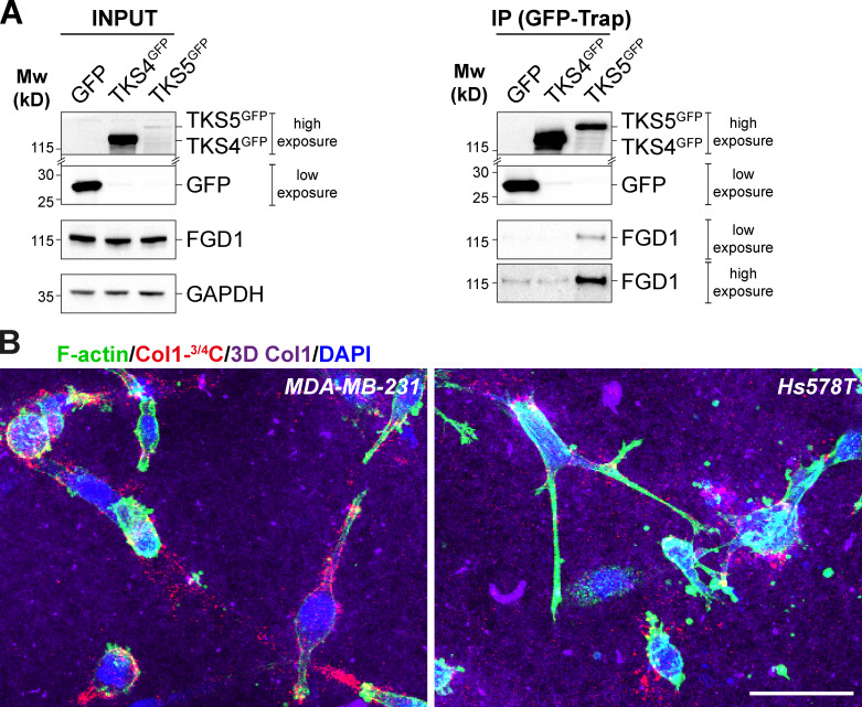
TKS4 does not interact with FGD1. (A) Lysates of MDA-MB-231 cells expressing TKS4GFP, TKS5GFP, or GFP (Ctrl) were immunoprecipitated with GFP antibodies. Bound proteins were analyzed with TKS4, TKS5, GFP, and FGD1 antibodies (GFP-Trap). Note that TKS4GFP and TKS5GFP signals correspond to a longer exposure than that of the GFP signal. In addition, in the immunoprecipitated materials (GFP-Trap), two different exposure times are shown for the FGD1 signal, indicating that there is no detectable binding of FGD1 to TKS4GFP. 5% of total lysates was loaded as a control (input). Equal loading was controlled using GAPDH antibody. Molecular weights are in kD. (B) MDA-MB-231 or Hs578T cells were embedded in 3D type I collagen gel (magenta) for 16 h and then stained for cytoskeletal F-actin (green). Pericellular collagenolysis was detected with the Col1-3/4C antibody (red). Nuclei were stained with DAPI (blue). Scale bar, 50 µm.

