Figure 7.
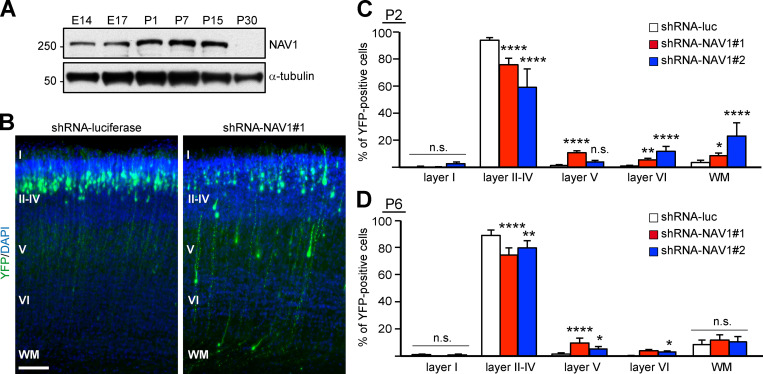
NAV1 participates in the radial migration of cortical projection neurons in the developing cerebral cortex. (A) Immunoblots of lysates of brain cortex at the indicated embryonic (E) and postnatal (P) days. α-Tubulin was used as a loading control. Results were replicated twice. (B) Coronal sections through the somatosensory cortex of P2 pups electroporated with the indicated shRNAs. DAPI was used for identification of cortical layers, labeled with roman numbers. WM, white matter. Scale bar, 100 µm. (C) Distribution of YFP-expressing cells in the indicated cortical layers from B. n = 4 (shRNA-luc), 3 (shRNA-NAV1#1), and 2 (shRNA-NAV1#2) mice. (D) Distribution of YFP-expressing cells in sections of P6 pups as in B. n = 4 mice per condition. Histograms show means ± SEM. Analyzed by χ2 test.

