Figure 3.
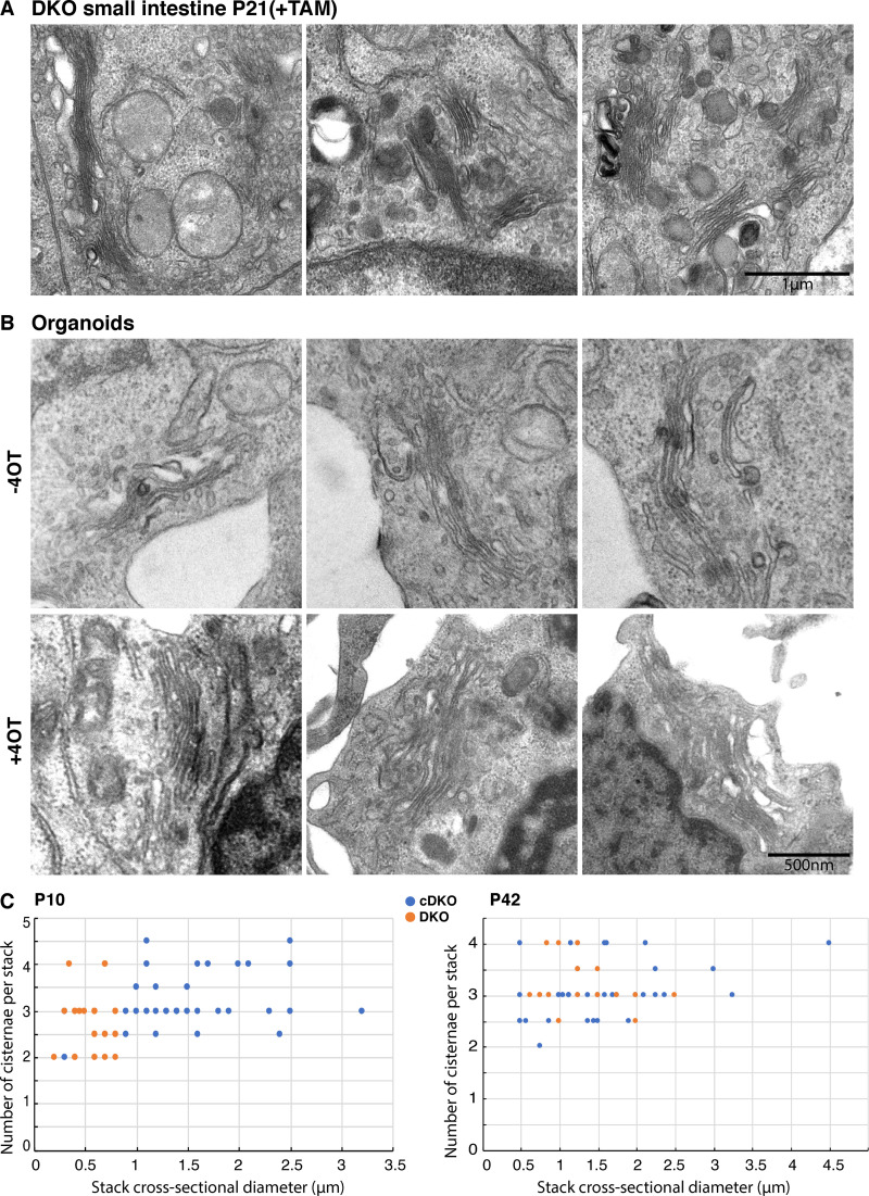
DKO of GORASP1 and GORASP2 does not lead to Golgi unstacking. (A) Electron micrograph of Golgi stacks in P21 small intestine from mouse treated with TAM at P1. Note that the Golgi are still stacked. (B) Electron micrograph of Golgi stacks in small intestine organoids treated with 4OT or without 4OT treatment for 7 d. Note that the Golgi are still stacked. (C) Quantification of the stack morphometry plotting the stack cross-sectional diameter versus the number of cisternae per stack in the small intestine of mice (±TAM at P1) at P10 (n = 35 and 29) and P42 (n = 40 and 20). Blue is the cDKO and orange the DKO. Note that the cross-sectional diameter is reduced upon TAM treatment but that the number of cisternae per stack is largely unchanged.

