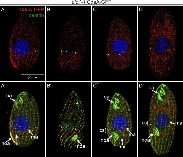
Figure 5.
The position of cortical subdivision correlates with the anterior margin of the posterior CdaA domain. (A–D′) Confocal images of elo1-1 mutant cells expressing CdaA-GFP stained with anti-GFP (red) and anti-centrin (green) antibodies and DAPI (blue). Examples of elo1-1 mutant cells before (A–B’) and during the formation of cortical subdivision (C–D’). (C–D′) Two elo1-1 cells with a cortical subdivision. The larger cell (D and D′) has a larger CdaA-GFP domain than the smaller cell (C and C′), but the cortical subdivision develops anteriorly to the anterior margin of CdaA-GFP. cs, cortical subdivision; mi, micronucleus; ma, macronucleus; noa, new oral apparatus (oral primordium); oa, oral apparatus. Arrowheads point to the anterior ends of some CdaA-GFP streaks.

