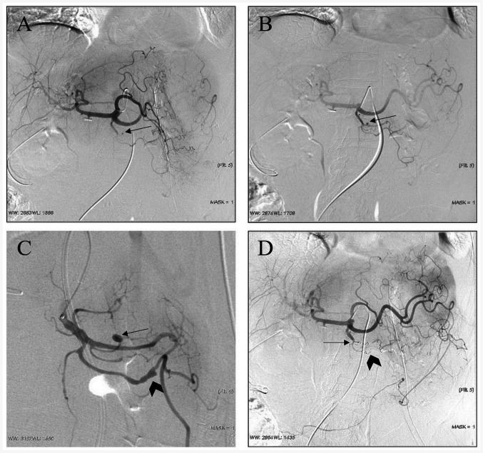
Figure 1.
Representative case of a 63-year-old female patient (case no. 1; Table I) diagnosed with duodenal adenocarcinoma. (A) Digital subtraction angiography images indicated a suspected pseudoaneurysm (arrow) in one branch of the gastroduodenal artery, which was confirmed as a saccular pseudoaneurysm (arrow) by (B) superselective catheterization to gastroduodenal arteriography. (C) Another irregular pseudoaneurysm is found by local magnification angiography (arrow). (D) A proximal embolization technique and exclusion technique were used, respectively, since the pseudoaneurysms arose from the end of a branch (arrow) and a branch with both inflow and outflow of the parental artery (arrowhead).

