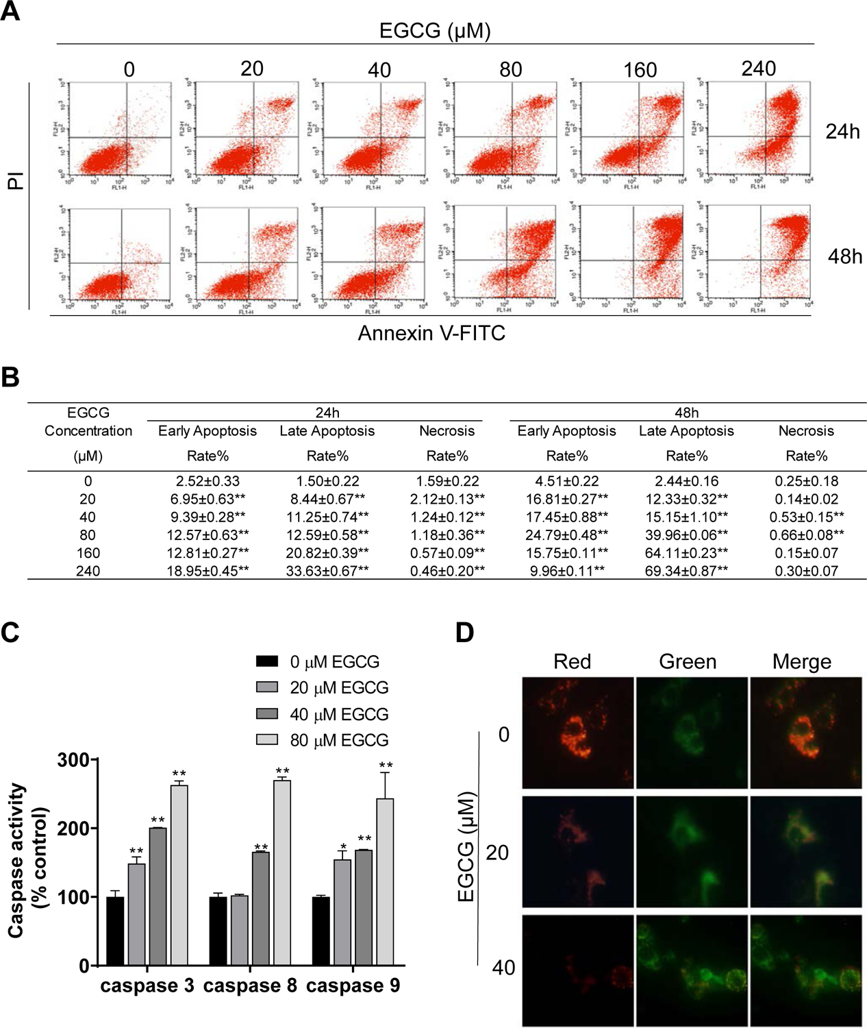
Figure 2: EGCG induced cell death by apoptosis in 4T1 breast cancer cells.

A: 4T1 cells treated with EGCG for 24 h and 48 h were stained with Annexin V/propidium iodide, and the percentage of apoptotic cells was determined by flow cytometry. B: Results are expressed as the mean ± SD. (*p<0.05, **p<0.01 vs. control). C: EGCG activated caspase activity in 4T1 cells. Results are expressed as percent control. (*p<0.05, **p<0.01 vs. control). D: EGCG promoted mitochondrial depolarization after 24 h incubation. Representative images (x400) of 4T1 cells treated with either vehicle (control) or EGCG and stained for JC-1 fluorescence.
