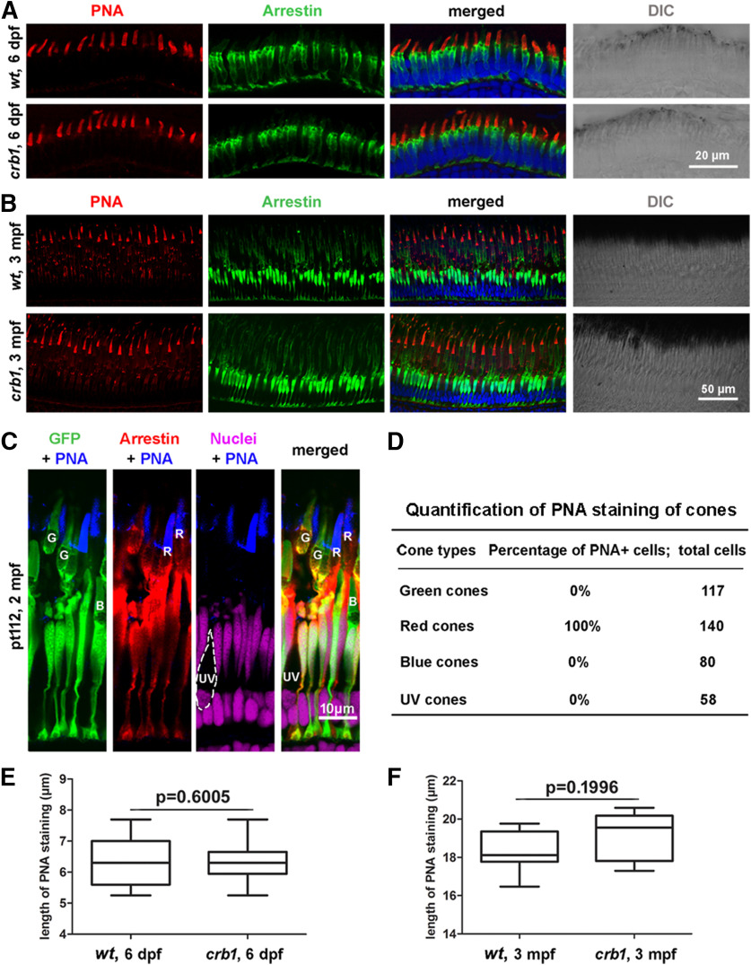
Figure 5.
The loss of Crb1 did not compromise photoreceptor morphologies at larval and adult stages. A, B, Morphologies of WT (wt) and crb1 mutant double cones, revealed by immunostaining of arrrestin3a with the Zpr1 antibody, and red cone outer segments, stained with PNA, at 6 dpf (A, scale bar, 20 μm) and 3 mpf (B, scale bar, 50 μm). DIC, Differential interference contrast. C, PNA-stained red cone outer segments. Cone types were determined by immunostaining of arrestin3a in double cones in the Tg (LCRRH2-RH2-1:GFP)pt112 transgenic background (pt112), which expresses GFP in green and blue cones (Fang et al., 2013). The following staining and morphologic features were used to distinguish four cone types: G, green cones, positive for both GFP and arrestin3a; R, red cones, negative for GFP but positive for arrestin3a; B, blue cones, positive for GFP positive but negative for arrestin3a; UV, UV cones, shortest cones with their outer segments closest to the OLM and negative for both GFP and arrestin3a. Scale bar, 10 μm. D, Quantification of PNA staining of four cone types. E, Quantification of the lengths of red cone outer segments of dark-adapted fish at 6 dpf (phenylthiourea-treated). Thirty cones from 6 WT and 41 cones from 6 crb1 mutant were quantified. Data are mean ± SEM. p value was generated by Student's t test, two-tailed hypothesis. F, Quantification of the lengths of red cone outer segments at the central retinal region of dark-adapted 3 mpf adult fish. Eighteen cones from 3 WT and 18 cones from 3 crb1 mutants were quantified. Data are mean ± SEM. p value was generated by Student's t test, two-tailed hypothesis.

