Figure 6.
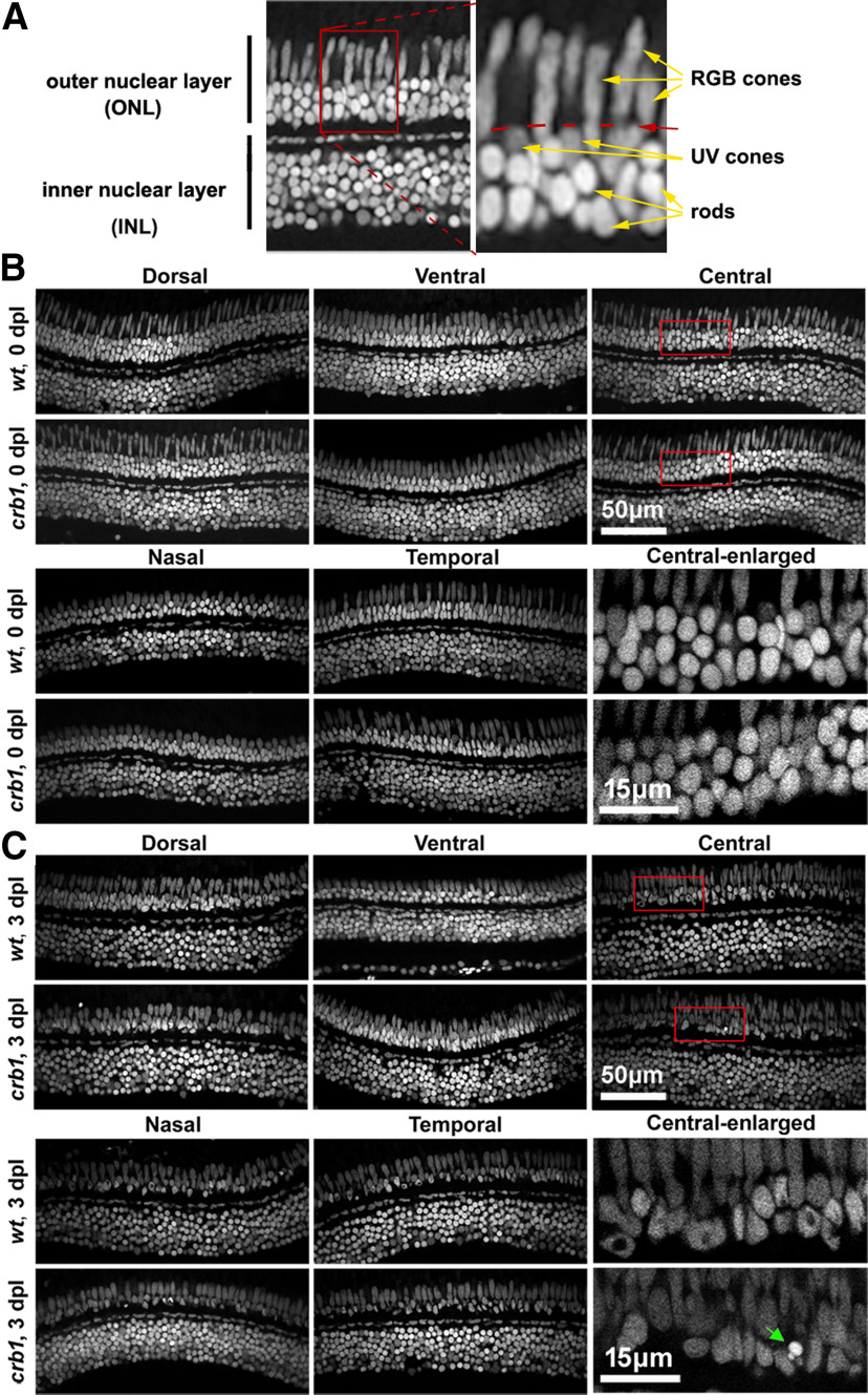
The loss of Crb1 made rod photoreceptors prone to damage by strong white light irradiation. A, An example DAPI image of a radial section of adult zebrafish retina, showing the outer nuclear layer (ONL) and the inner nuclear layer (INL). Arrows indicate the nuclei of RGB cones, UV cones, and rods. Red dashed lines indicate the position of the OLM. Left, Boxed region is magnified as the right panel. B, C, Typical images of radial retinal sections show the nuclear morphologies (by DAPI staining) of the ONL and INL of strong white light-treated fish at 0 dpl (B) and 3 dpl (C). B, C, Bottom right, Enlarged central retinal regions boxed in red. Green arrow indicates a highly condensed nuclear residue, possibly from a dead or dying rod. Scale bars, 50 μm or 15 μm.

