Figure 3.
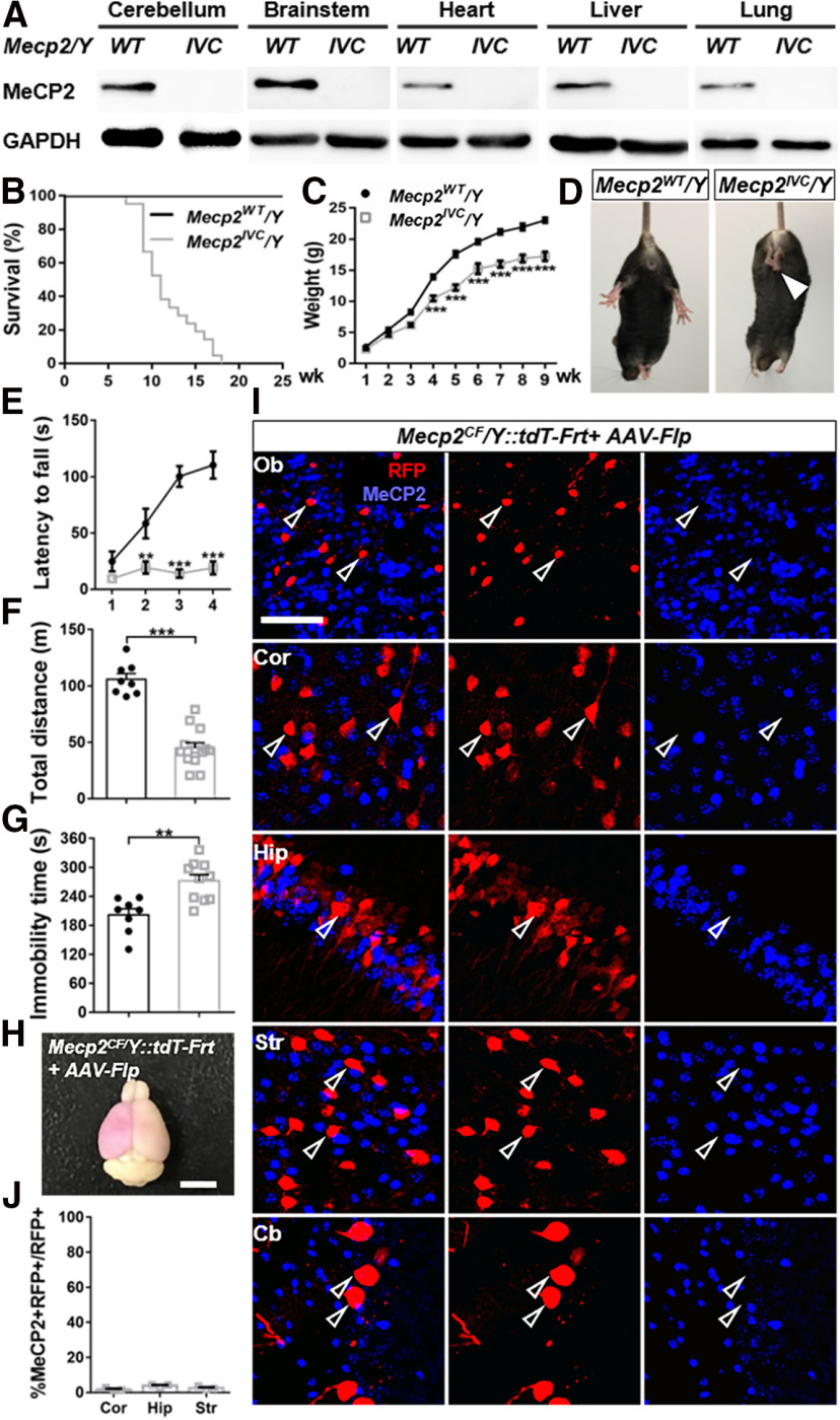
Efficient inactivation of Mecp2 by Cre- or Flp-mediated recombination. A, Western blots proving absence of full-length MeCP2 in all examined tissues in Mecp2IVC/Y. B, C, Reduced lifespan (B) and body weight (C) of Mecp2IVC/Y mice (survival curve, n = 27; body weight, n = 14) compared with Mecp2WT/Y mice (survival curve, n = 12; body weight, n = 8). D–G, Behavior deficits. D, Representative photographs of hindlimb clasping phenotype manifested in Mecp2IVC/Y. Arrowhead indicates clasping hindlimbs. E–G, Reduced motor coordination in rotarod test (E) and locomotor activity in open field (F) and increased immobility time in tail suspension (G) in Mecp2IVC/Y (n = 13, 13, 10) compared with Mecp2WT/Y (n = 9, 8, 8) littermates. H, Representative photograph of a brain from P0 AAV-Flp-injected, adult-perfused Mecp2CF/Y::tdT-Frt mouse. Purple, in bright-field image, represents higher-level RFP expression in a large number of infected cells in the brain. Scale bar, 500 μm. I, J, Efficient Mecp2 inactivation in AAV-Flp-infected cells in olfactory bulb (Ob), cortex (Cor), hippocampus (Hip), striatum (Str), and cerebellum (Cb). Scale bar, 50 μm. I, Representative images. J, Quantification. n = 3. Data are mean ± SEM. Dots and squares represent data from individual mice. n.s., Not significant at p > 0.05, **p < 0.01, ***p < 0.001. For more statistical details, see Table 2.

